4BQL image
Deposition Date 2013-05-31
Release Date 2014-02-12
Last Version Date 2024-05-08
Entry Detail
PDB ID:
4BQL
Title:
Crystal structure of archaeal actin
Biological Source:
Source Organism:
Host Organism:
Method Details:
Experimental Method:
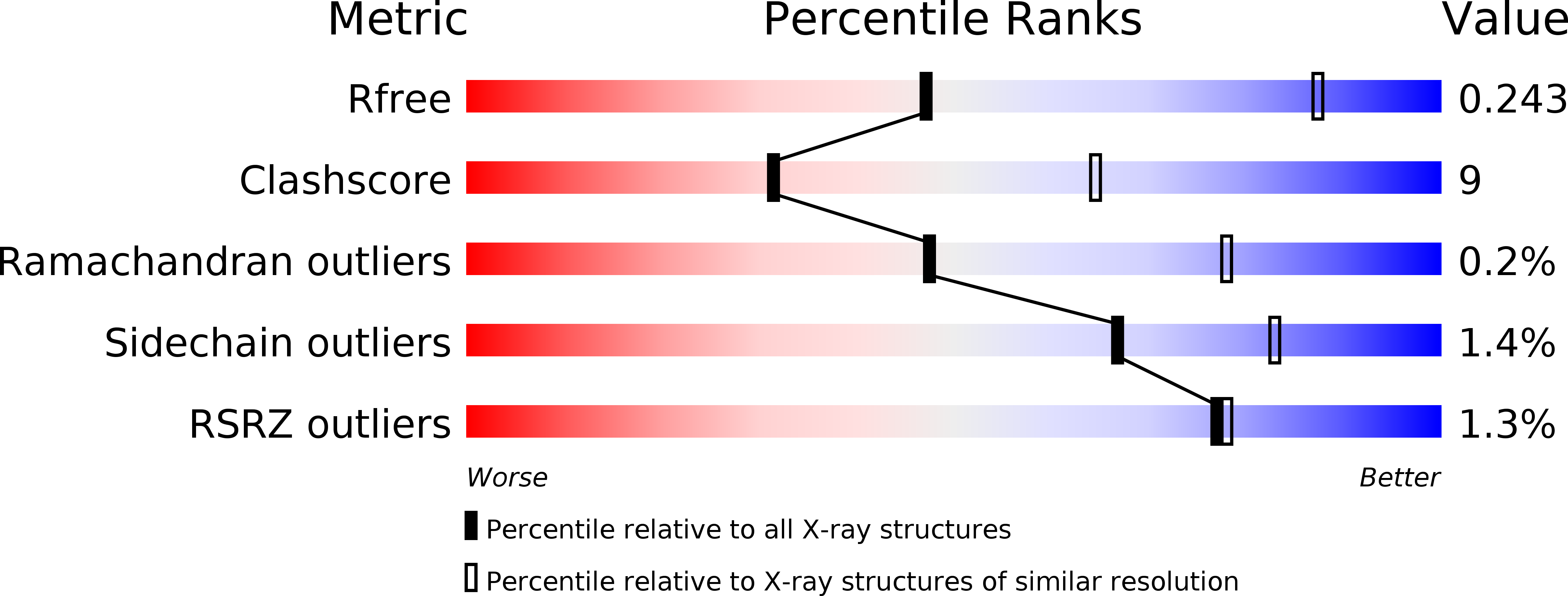
Resolution:
3.34 Å
R-Value Free:
0.24
R-Value Work:
0.20
R-Value Observed:
0.20
Space Group:
P 21 21 21
Macromolecular Entities
Polymer Type:polypeptide(L)
Molecule:ACTIN/ACTIN FAMILY PROTEIN
Gene (Uniprot):cren-1
Chain IDs:A, B, C, D
Chain Length:455
Number of Molecules:4
Biological Source:PYROBACULUM CALIDIFONTIS
Primary Citation
Structure of Crenactin, an Archaeal Actin Homologue Active at 90Degc.
Acta Crystallogr.,Sect.D 70 492 ? (2014)
PMID: 24531483 DOI: 10.1107/S1399004714000935

Abstact

The crystal structure of the archaeal actin, crenactin, from the rod-shaped hyperthermophilic (optimal growth at 90°C) crenarchaeon Pyrobaculum calidifontis is reported at 3.35 Å resolution. Despite low amino-acid sequence identity, the three-dimensional structure of the protein monomer is highly similar to those of eukaryotic actin and the bacterial MreB protein. Crenactin-specific features are also evident, as well as elements that are shared between crenactin and eukaryotic actin but are not found in MreB. In the crystal, crenactin monomers form right-handed helices, demonstrating that the protein is capable of forming filament-like structures. Monomer interactions in the helix, as well as interactions between crenactin and ADP in the nucleotide-binding pocket, are resolved at the atomic level and compared with those of actin and MreB. The results provide insights into the structural and functional properties of a heat-stable archaeal actin and contribute to the understanding of the evolution of actin-family proteins in the three domains of life.

Legend

Protein

Chemical

Disease

Primary Citation of related structures