4BQB image
Deposition Date 2013-05-30
Release Date 2013-06-12
Last Version Date 2024-11-06
Entry Detail
PDB ID:
4BQB
Keywords:
Title:
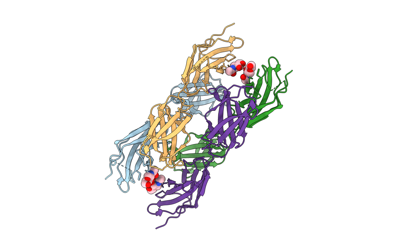
Crystal structure of the FN5 and FN6 domains of NEO1, form 2
Biological Source:
Source Organism:
MUS MUSCULUS (Taxon ID: 10090)
Host Organism:
Method Details:
Experimental Method:
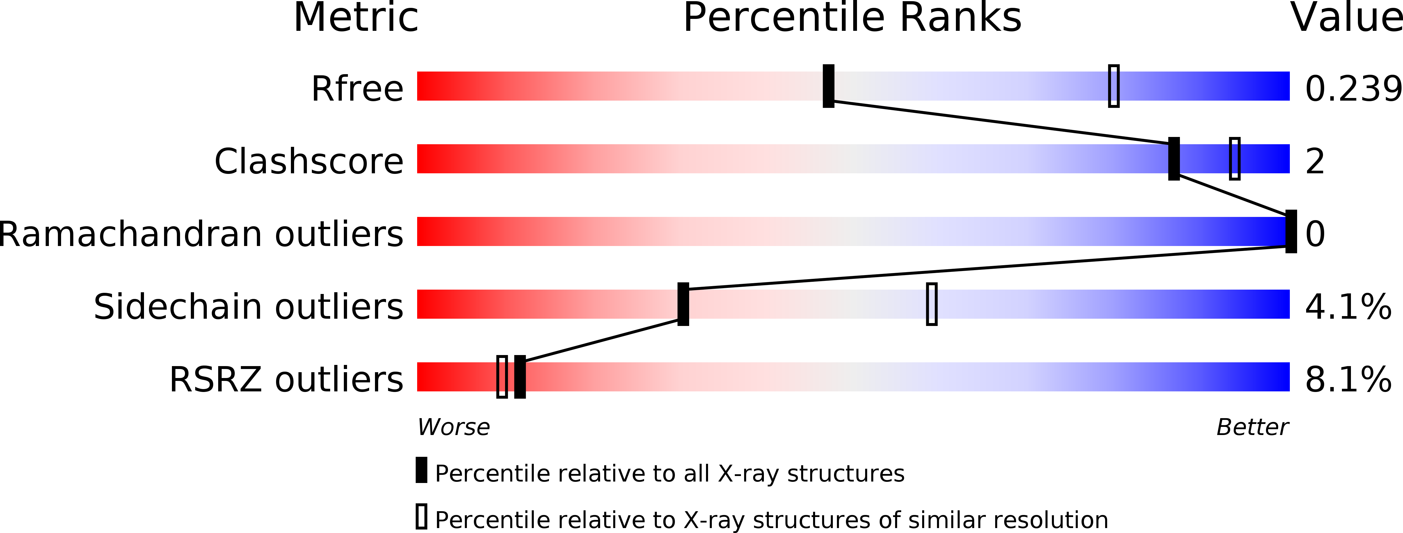
Resolution:
2.70 Å
R-Value Free:
0.22
R-Value Work:
0.19
R-Value Observed:
0.20
Space Group:
P 1 21 1
Macromolecular Entities
Structures with similar UniProt ID
Protein Blast
Polymer Type:polypeptide(L)
Molecule:NEOGENIN
Gene (Uniprot):Neo1
Chain IDs:A, B, C, D
Chain Length:264
Number of Molecules:4
Biological Source:MUS MUSCULUS
Modified Residue
Compound ID Chain ID Parent Comp ID Details 2D Image
ASN A ASN GLYCOSYLATION SITE
Ligand Molecules
Primary Citation
Structure of the Repulsive Guidance Molecule (Rgm)-Neogenin Signaling Hub
Science 341 77 ? (2013)
PMID: 23744777 DOI: 10.1126/SCIENCE.1232322

Abstact

Repulsive guidance molecule family members (RGMs) control fundamental and diverse cellular processes, including motility and adhesion, immune cell regulation, and systemic iron metabolism. However, it is not known how RGMs initiate signaling through their common cell-surface receptor, neogenin (NEO1). Here, we present crystal structures of the NEO1 RGM-binding region and its complex with human RGMB (also called dragon). The RGMB structure reveals a previously unknown protein fold and a functionally important autocatalytic cleavage mechanism and provides a framework to explain numerous disease-linked mutations in RGMs. In the complex, two RGMB ectodomains conformationally stabilize the juxtamembrane regions of two NEO1 receptors in a pH-dependent manner. We demonstrate that all RGM-NEO1 complexes share this architecture, which therefore represents the core of multiple signaling pathways.

Legend

Protein

Chemical

Disease

Primary Citation of related structures