4BPZ image
Deposition Date 2013-05-29
Release Date 2013-12-18
Last Version Date 2023-12-20
Entry Detail
PDB ID:
4BPZ
Keywords:
Title:
Crystal structure of lamA_E269S from Zobellia galactanivorans in complex with a trisaccharide of 1,3-1,4-beta-D-glucan.
Biological Source:
Source Organism:
Host Organism:
Method Details:
Experimental Method:
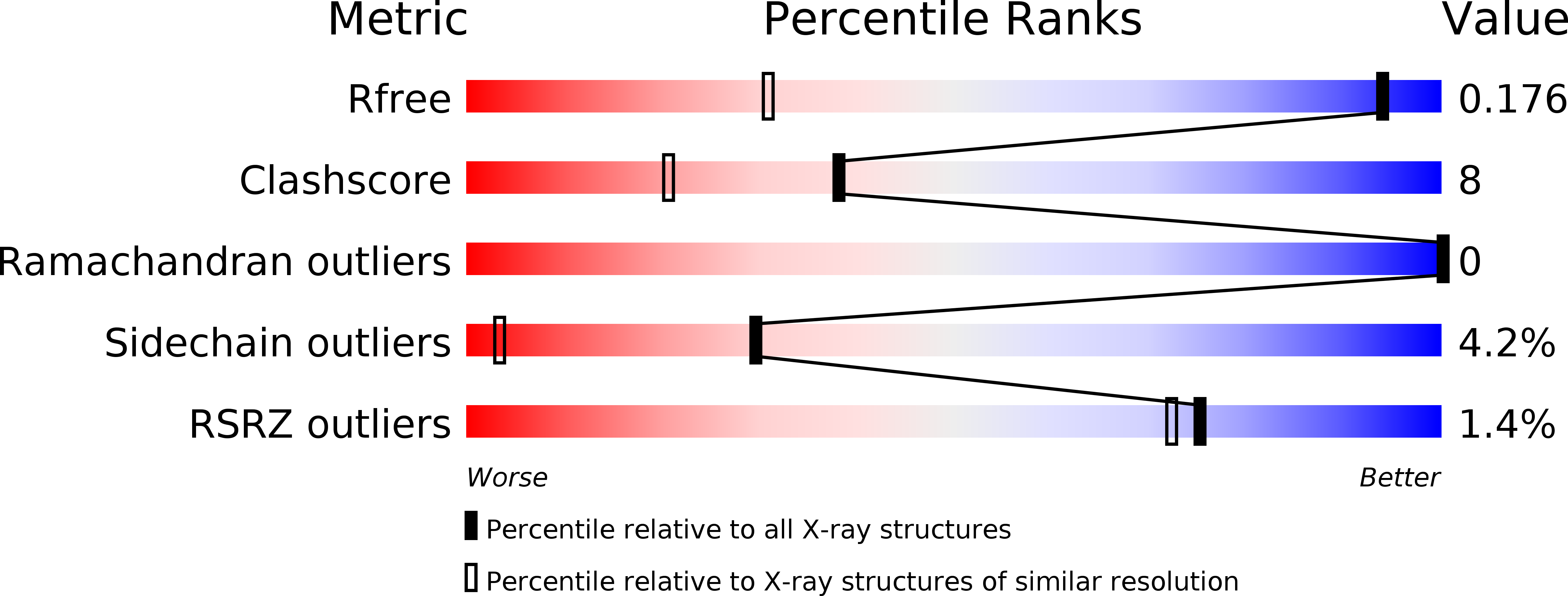
Resolution:
1.13 Å
R-Value Free:
0.17
R-Value Work:
0.14
R-Value Observed:
0.14
Space Group:
P 21 21 21
Macromolecular Entities
Structures with similar UniProt ID
Protein Blast
Polymer Type:polypeptide(L)
Molecule:ENDO-1,3-BETA-GLUCANASE, FAMILY GH16
Gene (Uniprot):lamA
Mutagens:YES
Chain IDs:A, B
Chain Length:256
Number of Molecules:2
Biological Source:ZOBELLIA GALACTANIVORANS
Ligand Molecules
Primary Citation
The Beta-Glucanase Zglama from Zobellia Galactanivorans Evolved a Bent Active Site Adapted for Efficient Degradation of Algal Laminarin.
J.Biol.Chem. 289 2027 ? (2014)
PMID: 24337571 DOI: 10.1074/JBC.M113.538843

Abstact

Laminarinase is commonly used to describe β-1,3-glucanases widespread throughout Archaea, bacteria, and several eukaryotic lineages. Some β-1,3-glucanases have already been structurally and biochemically characterized, but very few from organisms that are in contact with genuine laminarin, the storage polysaccharide of brown algae. Here we report the heterologous expression and subsequent biochemical and structural characterization of ZgLamAGH16 from Zobellia galactanivorans, the first GH16 laminarinase from a marine bacterium associated with seaweeds. ZgLamAGH16 contains a unique additional loop, compared with other GH16 laminarinases, which is composed of 17 amino acids and gives a bent shape to the active site cleft of the enzyme. This particular topology is perfectly adapted to the U-shaped conformation of laminarin chains in solution and thus explains the predominant specificity of ZgLamAGH16 for this substrate. The three-dimensional structure of the enzyme and two enzyme-substrate complexes, one with laminaritetraose and the other with a trisaccharide of 1,3-1,4-β-d-glucan, have been determined at 1.5, 1.35, and 1.13 Å resolution, respectively. The structural comparison of substrate recognition pattern between these complexes allows the proposition that ZgLamAGH16 likely diverged from an ancestral broad specificity GH16 β-glucanase and evolved toward a bent active site topology adapted to efficient degradation of algal laminarin.

Legend

Protein

Chemical

Disease

Primary Citation of related structures
Feedback Form
Name
Email
Institute
Feedback