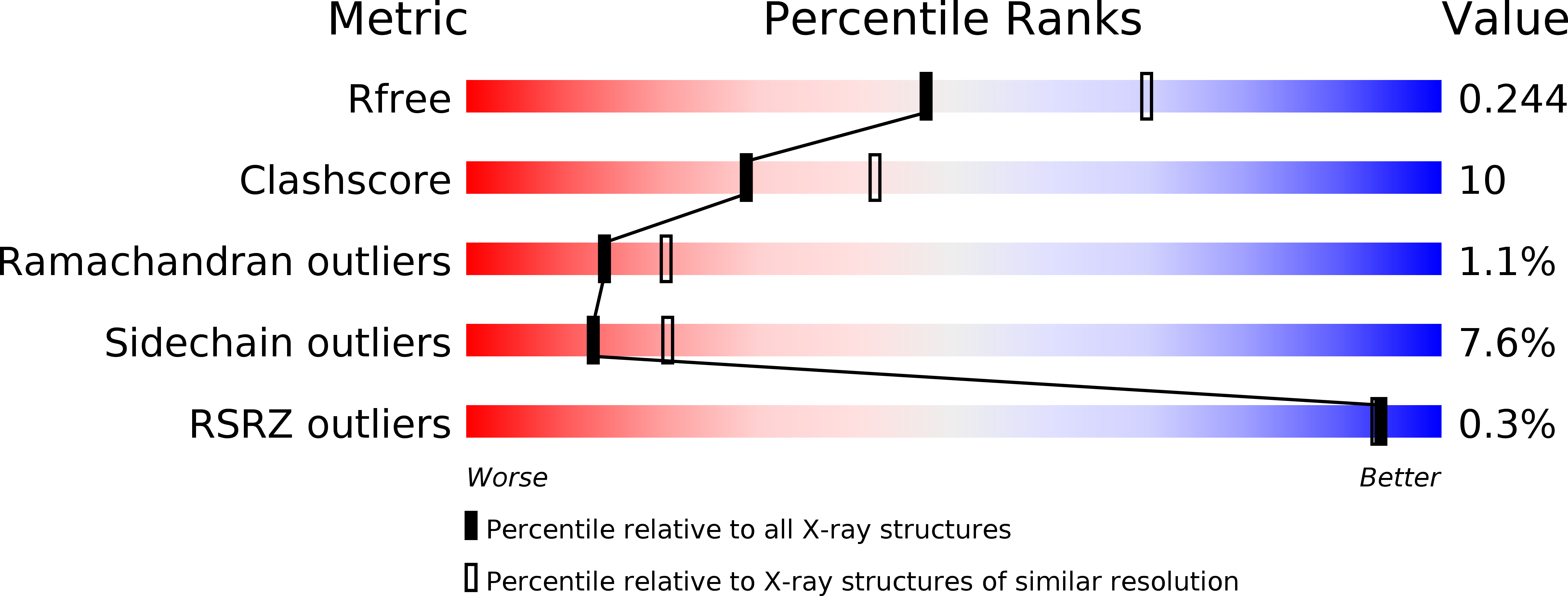
4BP8 image
Deposition Date 2013-05-23
Release Date 2014-02-12
Last Version Date 2024-10-23
Entry Detail
PDB ID:
4BP8
Keywords:
Title:
Oligopeptidase B from Trypanosoma brucei - open form
Biological Source:
Source Organism:
Host Organism:
Method Details:
Experimental Method:
Resolution:
2.40 Å
R-Value Free:
0.23
R-Value Work:
0.18
R-Value Observed:
0.18
Space Group:
P 32 2 1
Macromolecular Entities
Polymer Type:polypeptide(L)
Molecule:OLIGOPEPTIDASE B
Gene (Uniprot):opb
Chain IDs:A, B
Chain Length:715
Number of Molecules:2
Biological Source:TRYPANOSOMA BRUCEI
Modified Residue
Compound ID Chain ID Parent Comp ID Details 2D Image
MSE A MET SELENOMETHIONINE
Primary Citation
Crystal Structures of Trypanosoma Brucei Oligopeptidase B Broaden the Paradigm of Catalytic Regulation in Prolyl Oligopeptidase Family Enzymes.
Plos One 8 79349 ? (2013)
PMID: 24265767 DOI: 10.1371/JOURNAL.PONE.0079349

Abstact

Oligopeptidase B cleaves after basic amino acids in peptides up to 30 residues. As a virulence factor in bacteria and trypanosomatid pathogens that is absent in higher eukaryotes, this is a promising drug target. Here we present ligand-free open state and inhibitor-bound closed state crystal structures of oligopeptidase B from Trypanosoma brucei, the causative agent of African sleeping sickness. These (and related) structures show the importance of structural dynamics, governed by a fine enthalpic and entropic balance, in substrate size selectivity and catalysis. Peptides over 30 residues cannot fit the enzyme cavity, preventing the complete domain closure required for a key propeller Asp/Glu to fix the catalytic His and Arg in the catalytically competent conformation. This size exclusion mechanism protects larger peptides and proteins from degradation. Similar bacterial prolyl endopeptidase and archael acylaminoacyl peptidase structures demonstrate this mechanism is conserved among oligopeptidase family enzymes across all three domains of life.

Legend

Protein

Chemical

Disease

Primary Citation of related structures