Deposition Date
2013-04-21
Release Date
2013-06-12
Last Version Date
2024-05-08
Entry Detail
PDB ID:
4BK0
Keywords:
Title:
Crystal structure of the KIX domain of human RECQL5 (domain-swapped dimer)
Biological Source:
Source Organism(s):
HOMO SAPIENS (Taxon ID: 9606)
Expression System(s):
Method Details:
Experimental Method:
Resolution:
1.90 Å
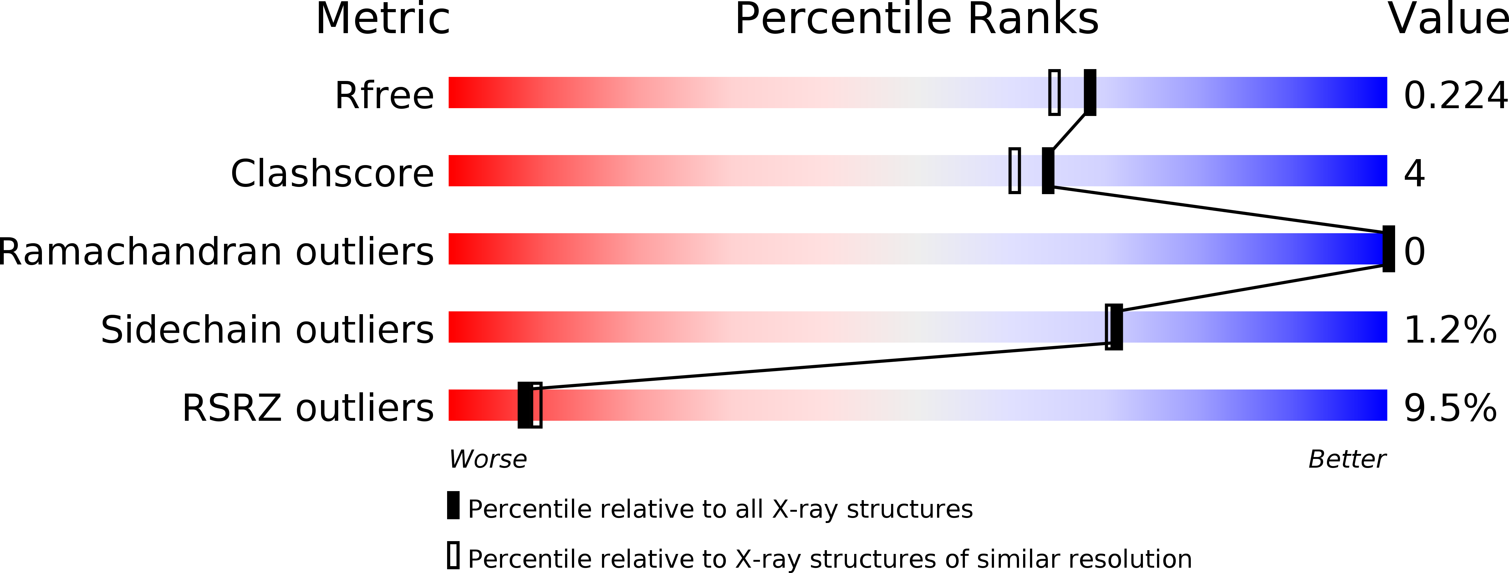
R-Value Free:
0.23
R-Value Work:
0.20
R-Value Observed:
0.20
Space Group:
P 21 21 21