4BGF image
Entry Detail
PDB ID:
4BGF
Keywords:
Title:
The 3D-structure of arylamine-N-acetyltransferase from M. tuberculosis
Biological Source:
Host Organism:
PDB Version:
Deposition Date:
2013-03-26
Release Date:
2013-07-24
Method Details:
Experimental Method:
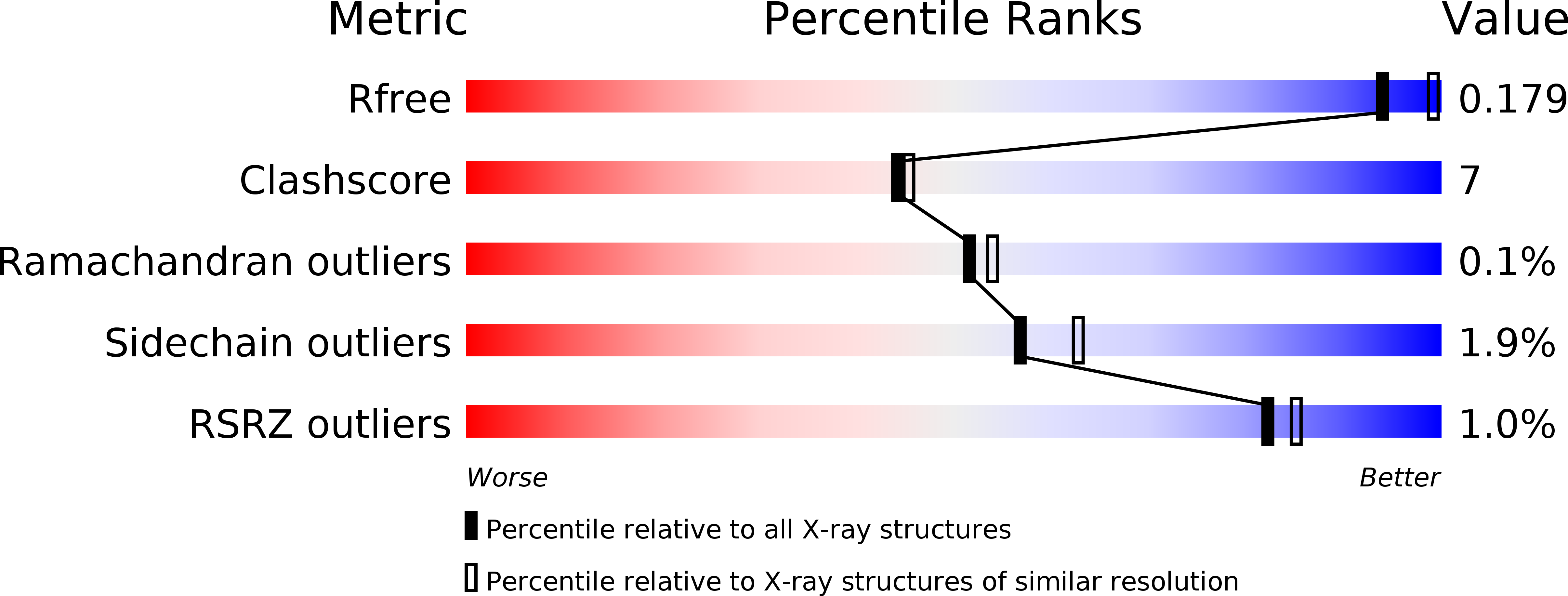
Resolution:
2.10 Å
R-Value Free:
0.18
R-Value Work:
0.16
R-Value Observed:
0.16
Space Group:
P 1 21 1
Macromolecular Entities
Polymer Type:polypeptide(L)
Description:ARYLAMINE N-ACETYLTRANSFERASE NAT
Chain IDs:A, B, C, D, E, F, G, H
Chain Length:283
Number of Molecules:8
Biological Source:MYCOBACTERIUM TUBERCULOSIS
Primary Citation
Structure of arylamine N-acetyltransferase from Mycobacterium tuberculosis determined by cross-seeding with the homologous protein from M. marinum: triumph over adversity.
Acta Crystallogr. D Biol. Crystallogr. 69 1433 1446 (2013)
PMID: 23897467 DOI: 10.1107/S0907444913015126

Abstact

Arylamine N-acetyltransferase from Mycobacterium tuberculosis (TBNAT) plays an important role in the intracellular survival of the microorganism inside macrophages. Medicinal chemistry efforts to optimize inhibitors of the TBNAT enzyme have been hampered by the lack of a three-dimensional structure of the enzyme. In this paper, the first structure of TBNAT, determined using a lone crystal produced using cross-seeding with the homologous protein from M. marinum, is reported. Despite the similarity between the two enzymes (74% sequence identity), they show distinct physical and biochemical characteristics. The structure elegantly reveals the characteristic features of the protein surface as well as details of the active site of TBNAT relevant to drug-discovery efforts. The crystallographic analysis of the diffraction data presented many challenges, since the crystal was twinned and the habit possessed pseudo-translational symmetry.

Legend

Protein

Chemical

Disease

Primary Citation of related structures