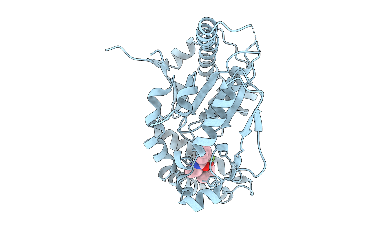
4BFS image
Entry Detail
PDB ID:
4BFS
Keywords:
Title:
Crystal structure of Mycobacterium tuberculosis PanK in complex with a triazole inhibitory compound (1a)
Biological Source:
Host Organism:
PDB Version:
Deposition Date:
2013-03-22
Release Date:
2013-05-15
Method Details:
Experimental Method:
Resolution:
2.90 Å
R-Value Free:
0.24
R-Value Work:
0.19
R-Value Observed:
0.19
Space Group:
P 31 2 1
Macromolecular Entities
Polymer Type:polypeptide(L)
Description:PANTOTHENATE KINASE
Chain IDs:A
Chain Length:318
Number of Molecules:1
Biological Source:MYCOBACTERIUM TUBERCULOSIS
Ligand Molecules
Primary Citation
Structural and Biochemical Characterization of Compounds Inhibiting Mycobacterium Tuberculosis Pank
J.Biol.Chem. 288 18260 ? (2013)
PMID: 23661699 DOI: 10.1074/JBC.M113.476473

Abstact

Mycobacterium tuberculosis, the bacterial causative agent of tuberculosis, currently affects millions of people. The emergence of drug-resistant strains makes development of new antibiotics targeting the bacterium a global health priority. Pantothenate kinase, a key enzyme in the universal biosynthesis of the essential cofactor CoA, was targeted in this study to find new tuberculosis drugs. The biochemical characterizations of two new classes of compounds that inhibit pantothenate kinase from M. tuberculosis are described, along with crystal structures of their enzyme-inhibitor complexes. These represent the first crystal structures of this enzyme with engineered inhibitors. Both classes of compounds bind in the active site of the enzyme, overlapping with the binding sites of the natural substrate and product, pantothenate and phosphopantothenate, respectively. One class of compounds also interferes with binding of the cofactor ATP. The complexes were crystallized in two crystal forms, one of which is in a new space group for this enzyme and diffracts to the highest resolution reported for any pantothenate kinase structure. These two crystal forms allowed, for the first time, modeling of the cofactor-binding loop in both open and closed conformations. The structures also show a binding mode of ATP different from that previously reported for the M. tuberculosis enzyme but similar to that in the pantothenate kinases of other organisms.

Legend

Protein

Chemical

Disease

Primary Citation of related structures