
Deposition Date
2012-10-08
Release Date
2013-05-15
Last Version Date
2024-10-23
Entry Detail
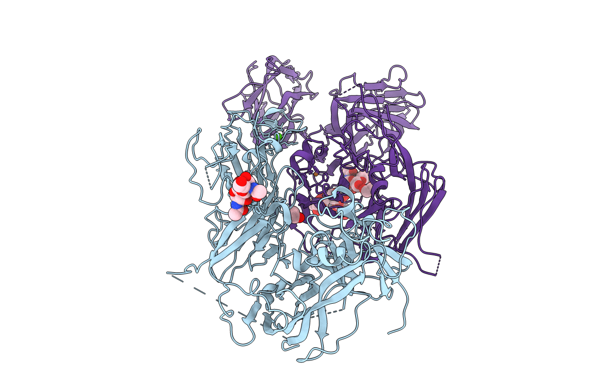
PDB ID:
4BDV
Keywords:
Title:
CRYSTAL STRUCTURE OF A TRUNCATED B-DOMAIN HUMAN FACTOR VIII
Biological Source:
Source Organism(s):
HOMO SAPIENS (Taxon ID: 9606)
Expression System(s):
Method Details:
Experimental Method:
Resolution:
3.98 Å
R-Value Free:
0.24
R-Value Work:
0.16
R-Value Observed:
0.16
Space Group:
P 41 21 2
