4BC5 image
Deposition Date 2012-10-01
Release Date 2012-11-28
Last Version Date 2024-05-01
Entry Detail
PDB ID:
4BC5
Keywords:
Title:
Crystal structure of human D-xylulokinase in complex with inhibitor 5- deoxy-5-fluoro-D-xylulose
Biological Source:
Source Organism:
HOMO SAPIENS (Taxon ID: 9606)
Host Organism:
Method Details:
Experimental Method:
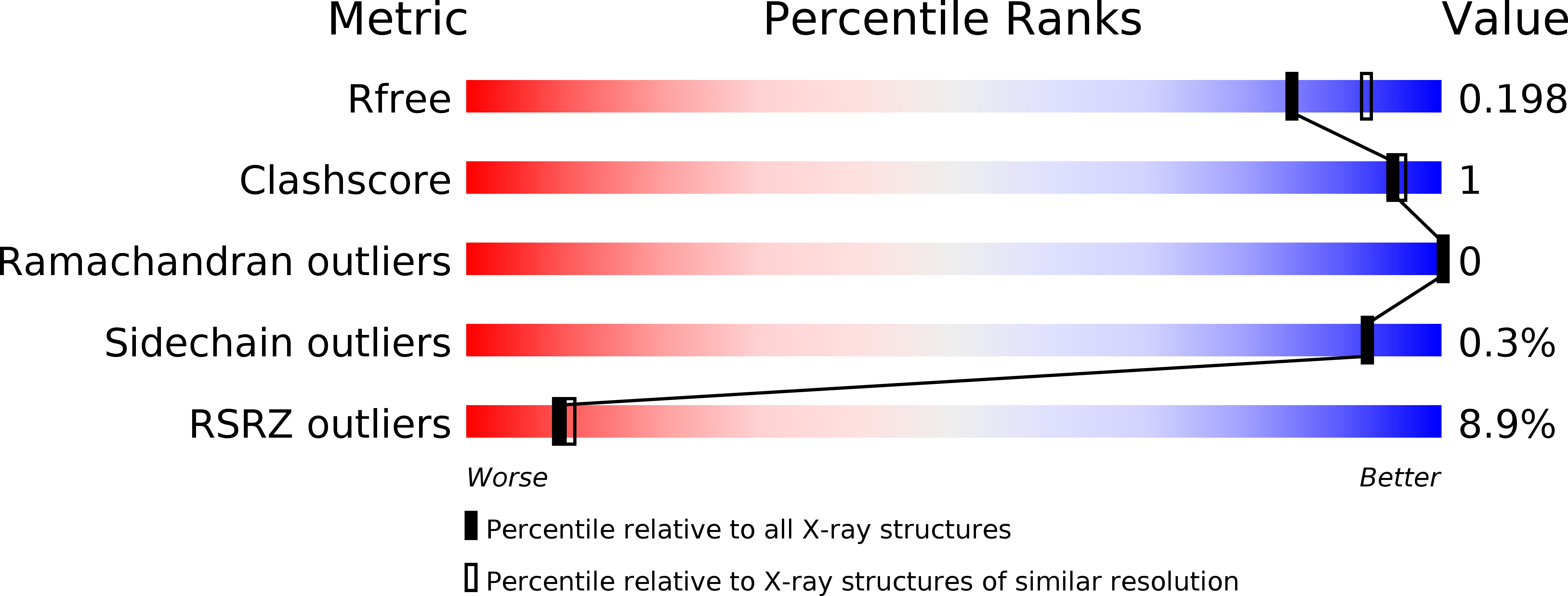
Resolution:
1.98 Å
R-Value Free:
0.19
R-Value Work:
0.17
R-Value Observed:
0.17
Space Group:
P 32
Macromolecular Entities
Structures with similar UniProt ID
Protein Blast
Polymer Type:polypeptide(L)
Molecule:XYLULOSE KINASE
Gene (Uniprot):XYLB
Chain IDs:A, B, C
Chain Length:538
Number of Molecules:3
Biological Source:HOMO SAPIENS
Primary Citation
Structure and Function of Human Xylulokinase, an Enzyme with Important Roles in Carbohydrate Metabolism
J.Biol.Chem. 288 1643 ? (2013)
PMID: 23179721 DOI: 10.1074/JBC.M112.427997

Abstact

D-Xylulokinase (XK; EC 2.7.1.17) catalyzes the ATP-dependent phosphorylation of d-xylulose (Xu) to produce xylulose 5-phosphate (Xu5P). In mammals, XK is the last enzyme in the glucuronate-xylulose pathway, active in the liver and kidneys, and is linked through its product Xu5P to the pentose-phosphate pathway. XK may play an important role in metabolic disease, given that Xu5P is a key regulator of glucose metabolism and lipogenesis. We have expressed the product of a putative human XK gene and identified it as the authentic human d-xylulokinase (hXK). NMR studies with a variety of sugars showed that hXK acts only on d-xylulose, and a coupled photometric assay established its key kinetic parameters as K(m)(Xu) = 24 ± 3 μm and k(cat) = 35 ± 5 s(-1). Crystal structures were determined for hXK, on its own and in complexes with Xu, ADP, and a fluorinated inhibitor. These reveal that hXK has a two-domain fold characteristic of the sugar kinase/hsp70/actin superfamily, with glycerol kinase as its closest relative. Xu binds to domain-I and ADP to domain-II, but in this open form of hXK they are 10 Å apart, implying that a large scale conformational change is required for catalysis. Xu binds in its linear keto-form, sandwiched between a Trp side chain and polar side chains that provide exquisite hydrogen bonding recognition. The hXK structure provides a basis for the design of specific inhibitors with which to probe its roles in sugar metabolism and metabolic disease.

Legend

Protein

Chemical

Disease

Primary Citation of related structures
Feedback Form
Name
Email
Institute
Feedback