
Deposition Date
2012-09-28
Release Date
2012-11-14
Last Version Date
2025-04-09
Entry Detail
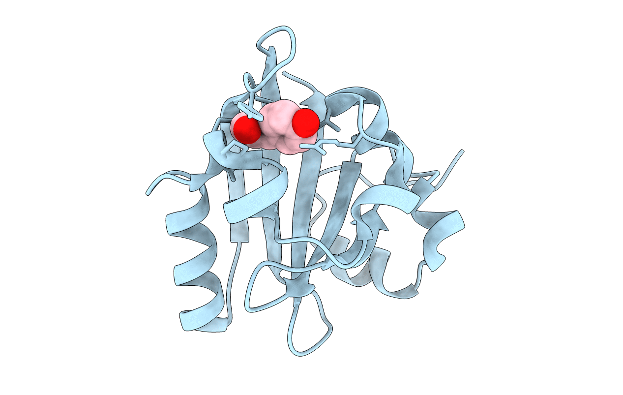
PDB ID:
4BBV
Keywords:
Title:
The PB0 Photocycle Intermediate of Photoactive Yellow Protein
Biological Source:
Source Organism(s):
HALORHODOSPIRA HALOPHILA (Taxon ID: 1053)
Expression System(s):
Method Details:
Experimental Method:
Resolution:
1.60 Å
R-Value Free:
0.36
R-Value Work:
0.25
R-Value Observed:
0.25
Space Group:
P 63
