4B9Q image
Deposition Date 2012-09-06
Release Date 2012-11-14
Last Version Date 2024-11-06
Entry Detail
PDB ID:
4B9Q
Keywords:
Title:
Open conformation of ATP-bound Hsp70 homolog DnaK
Biological Source:
Source Organism:
ESCHERICHIA COLI (Taxon ID: 562)
Host Organism:
Method Details:
Experimental Method:
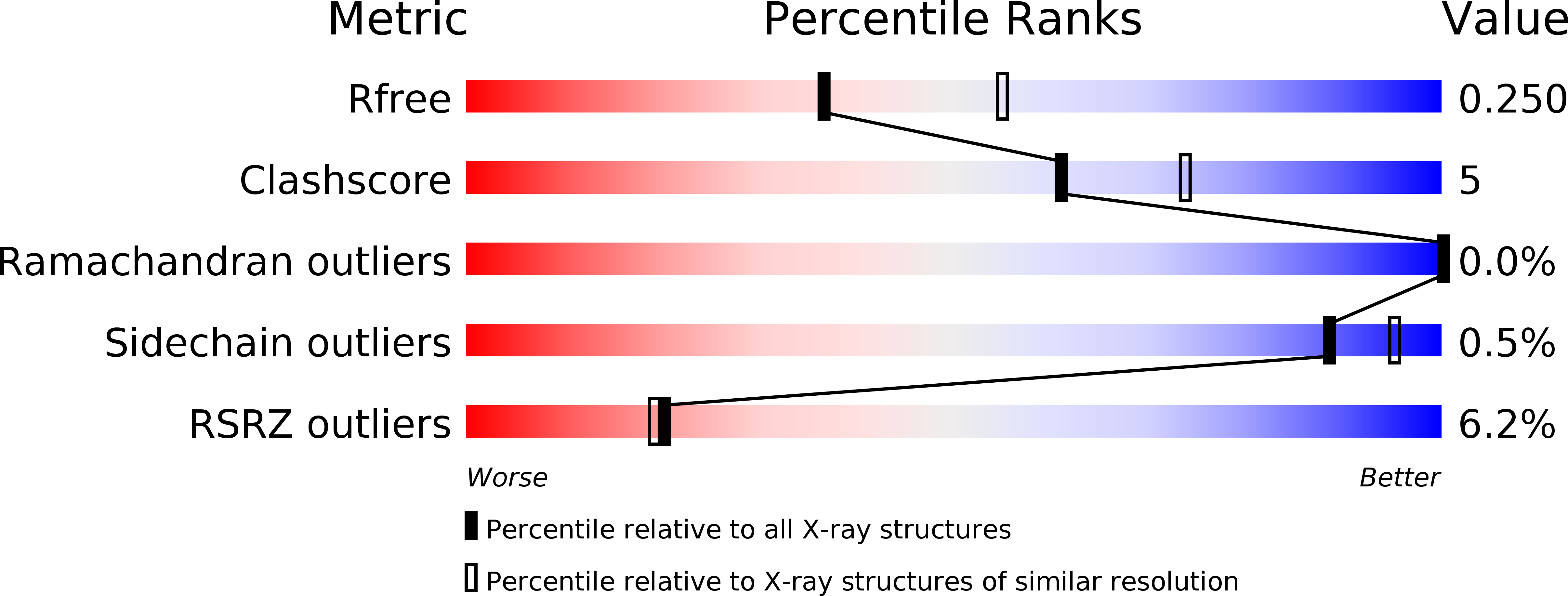
Resolution:
2.40 Å
R-Value Free:
0.23
R-Value Work:
0.19
R-Value Observed:
0.19
Space Group:
C 1 2 1
Macromolecular Entities
Polymer Type:polypeptide(L)
Molecule:CHAPERONE PROTEIN DNAK
Gene (Uniprot):dnaK
Mutations:YES
Chain IDs:A, B, C, D
Chain Length:605
Number of Molecules:4
Biological Source:ESCHERICHIA COLI
Modified Residue
Compound ID Chain ID Parent Comp ID Details 2D Image
MSE A MET SELENOMETHIONINE
Primary Citation
Structure and Dynamics of the ATP-Bound Open Conformation of Hsp70 Chaperones
Mol.Cell 48 863 ? (2012)
PMID: 23123194 DOI: 10.1016/J.MOLCEL.2012.09.023

Abstact

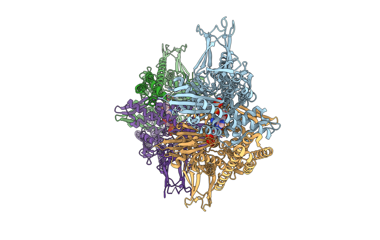
Central to the chaperone function of Hsp70s is the transition between open and closed conformations of their polypeptide substrate binding domain (SBD), which is regulated through an allosteric mechanism via ATP binding and hydrolysis in their nucleotide binding domain (NBD). Although the structure of the closed conformation of Hsp70s is well studied, the open conformation has remained elusive. Here, we report on the 2.4 Å crystal structure of the ATP-bound open conformation of the Escherichia coli Hsp70 homolog DnaK. In the open DnaK structure, the β sheet and α-helical lid subdomains of the SBD are detached from one another and docked to different faces of the NBD. The contacts between the β sheet subdomain and the NBD reveal the mechanism of allosteric regulation. In addition, we demonstrate that docking of the β sheet and α-helical lid subdomains to the NBD is a sequential process influenced by peptide and protein substrates.

Legend

Protein

Chemical

Disease

Primary Citation of related structures