4B5Q image
Deposition Date 2012-08-07
Release Date 2013-04-03
Last Version Date 2024-10-23
Entry Detail
PDB ID:
4B5Q
Keywords:
Title:
The lytic polysaccharide monooxygenase GH61D structure from the basidiomycota fungus Phanerochaete chrysosporium
Biological Source:
Source Organism:
Host Organism:
Method Details:
Experimental Method:
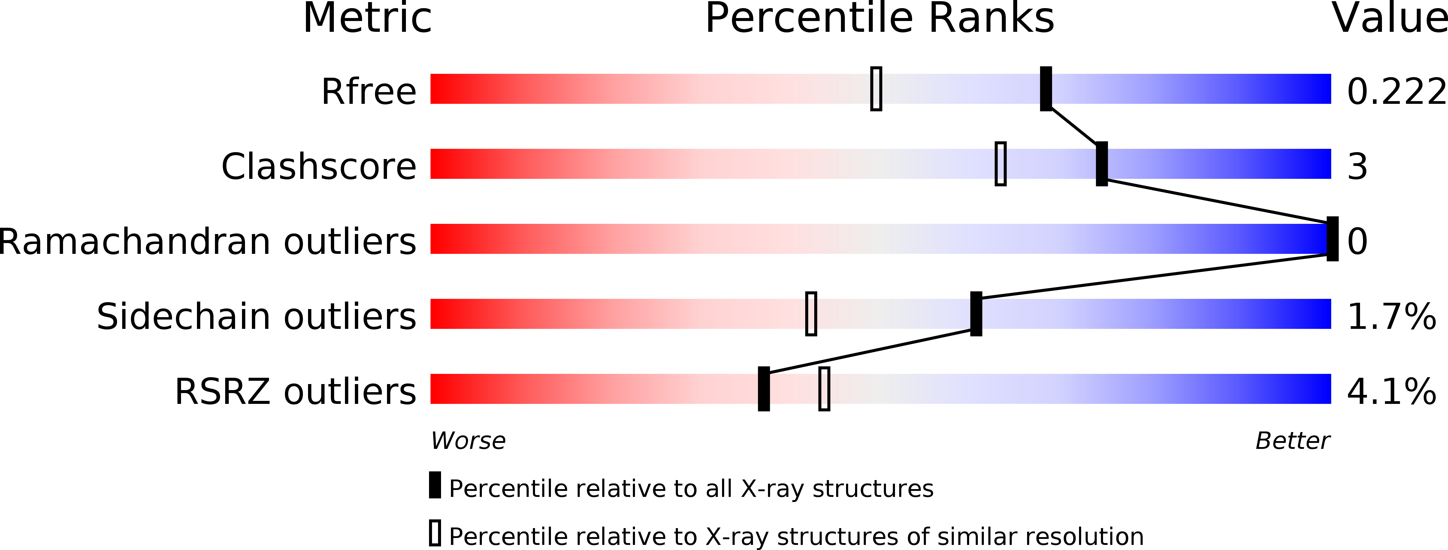
Resolution:
1.75 Å
R-Value Free:
0.22
R-Value Work:
0.18
R-Value Observed:
0.18
Space Group:
C 1 2 1
Macromolecular Entities
Structures with similar UniProt ID
Protein Blast
Polymer Type:polypeptide(L)
Molecule:GLYCOSIDE HYDROLASE FAMILY 61 PROTEIN D
Gene (Uniprot):LPMO9D
Chain IDs:A, B
Chain Length:217
Number of Molecules:2
Biological Source:PHANEROCHAETE CHRYSOSPORIUM
Modified Residue
Compound ID Chain ID Parent Comp ID Details 2D Image
SER A SER GLYCOSYLATION SITE
Primary Citation
Crystal Structure and Computational Characterization of the Lytic Polysaccharide Monooxygenase Gh61D from the Basidiomycota Fungus Phanerochaete Chrysosporium
J.Biol.Chem. 288 12828 ? (2013)
PMID: 23525113 DOI: 10.1074/JBC.M113.459396

Abstact

Carbohydrate structures are modified and degraded in the biosphere by a myriad of mostly hydrolytic enzymes. Recently, lytic polysaccharide mono-oxygenases (LPMOs) were discovered as a new class of enzymes for cleavage of recalcitrant polysaccharides that instead employ an oxidative mechanism. LPMOs employ copper as the catalytic metal and are dependent on oxygen and reducing agents for activity. LPMOs are found in many fungi and bacteria, but to date no basidiomycete LPMO has been structurally characterized. Here we present the three-dimensional crystal structure of the basidiomycete Phanerochaete chrysosporium GH61D LPMO, and, for the first time, measure the product distribution of LPMO action on a lignocellulosic substrate. The structure reveals a copper-bound active site common to LPMOs, a collection of aromatic and polar residues near the binding surface that may be responsible for regio-selectivity, and substantial differences in loop structures near the binding face compared with other LPMO structures. The activity assays indicate that this LPMO primarily produces aldonic acids. Last, molecular simulations reveal conformational changes, including the binding of several regions to the cellulose surface, leading to alignment of three tyrosine residues on the binding face of the enzyme with individual cellulose chains, similar to what has been observed for family 1 carbohydrate-binding modules. A calculated potential energy surface for surface translation indicates that P. chrysosporium GH61D exhibits energy wells whose spacing seems adapted to the spacing of cellobiose units along a cellulose chain.

Legend

Protein

Chemical

Disease

Primary Citation of related structures