4B5P image
Deposition Date 2012-08-07
Release Date 2012-10-24
Last Version Date 2023-12-20
Entry Detail
PDB ID:
4B5P
Keywords:
Title:
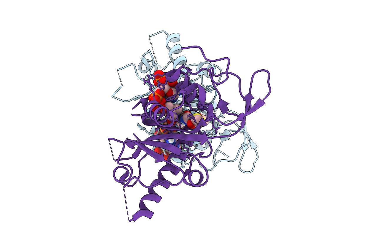
Crystal structure of human alpha tubulin acetyltransferase catalytic domain Q58A variant
Biological Source:
Source Organism:
HOMO SAPIENS (Taxon ID: 9606)
Host Organism:
Method Details:
Experimental Method:
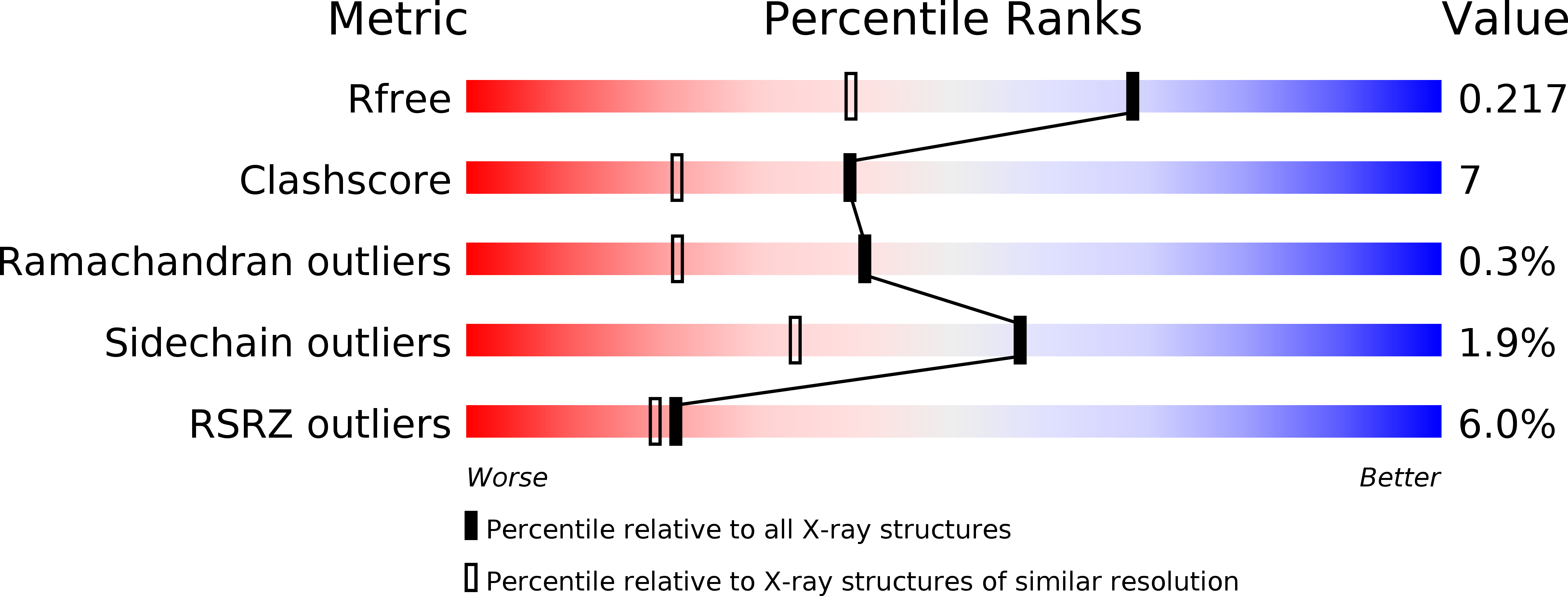
Resolution:
1.60 Å
R-Value Free:
0.22
R-Value Work:
0.18
R-Value Observed:
0.18
Space Group:
P 21 21 21
Macromolecular Entities
Structures with similar UniProt ID
Protein Blast
Polymer Type:polypeptide(L)
Molecule:ALPHA-TUBULIN N-ACETYLTRANSFERASE
Gene (Uniprot):ATAT1
Mutagens:YES
Chain IDs:A
Chain Length:200
Number of Molecules:1
Biological Source:HOMO SAPIENS
Structures with similar UniProt ID
Protein Blast
Polymer Type:polypeptide(L)
Molecule:ALPHA-TUBULIN N-ACETYLTRANSFERASE
Gene (Uniprot):ATAT1
Mutagens:YES
Chain IDs:B
Chain Length:200
Number of Molecules:1
Biological Source:HOMO SAPIENS
Primary Citation
Atomic Resolution Structure of Human Alpha-Tubulin Acetyltransferase Bound to Acetyl-Coa.
Proc.Natl.Acad.Sci.USA 109 19649 ? (2012)
PMID: 23071318 DOI: 10.1073/PNAS.1209343109

Abstact

Acetylation of lysine residues is an important posttranslational modification found in all domains of life. α-Tubulin is specifically acetylated on lysine 40, a modification that serves to stabilize microtubules of axons and cilia. Whereas histone acetyltransferases have been extensively studied, there is no structural and mechanistic information available on α-tubulin acetyltransferases. Here, we present the structure of the human α-tubulin acetyltransferase catalytic domain bound to its cosubstrate acetyl-CoA at 1.05 Å resolution. Compared with other lysine acetyltransferases of known structure, α-tubulin acetyltransferase displays a relatively well-conserved cosubstrate binding pocket but is unique in its active site and putative α-tubulin binding site. Using acetylation assays with structure-guided mutants, we map residues important for acetyl-CoA binding, substrate binding, and catalysis. This analysis reveals a basic patch implicated in substrate binding and a conserved glutamine residue required for catalysis, demonstrating that the family of α-tubulin acetyltransferases uses a reaction mechanism different from other lysine acetyltransferases characterized to date.

Legend

Protein

Chemical

Disease

Primary Citation of related structures