
Deposition Date
2012-07-12
Release Date
2013-07-31
Last Version Date
2023-12-20
Entry Detail
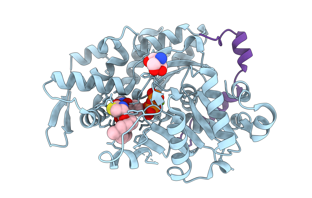
PDB ID:
4B1U
Keywords:
Title:
Structure of the Phactr1 RPEL domain and RPEL motif directed assemblies with G-actin reveal the molecular basis for actin binding cooperativity.
Biological Source:
Source Organism(s):
ORYCTOLAGUS CUNICULUS (Taxon ID: 9986)
Method Details:
Experimental Method:
Resolution:
2.00 Å
R-Value Free:
0.23
R-Value Work:
0.19
R-Value Observed:
0.19
Space Group:
P 32 2 1
