Deposition Date
2012-06-25
Release Date
2012-08-08
Last Version Date
2024-11-20
Entry Detail
PDB ID:
4AZA
Keywords:
Title:
Improved eIF4E binding peptides by phage display guided design.
Biological Source:
Source Organism(s):
HOMO SAPIENS (Taxon ID: 9606)
Expression System(s):
Method Details:
Experimental Method:
Resolution:
2.16 Å
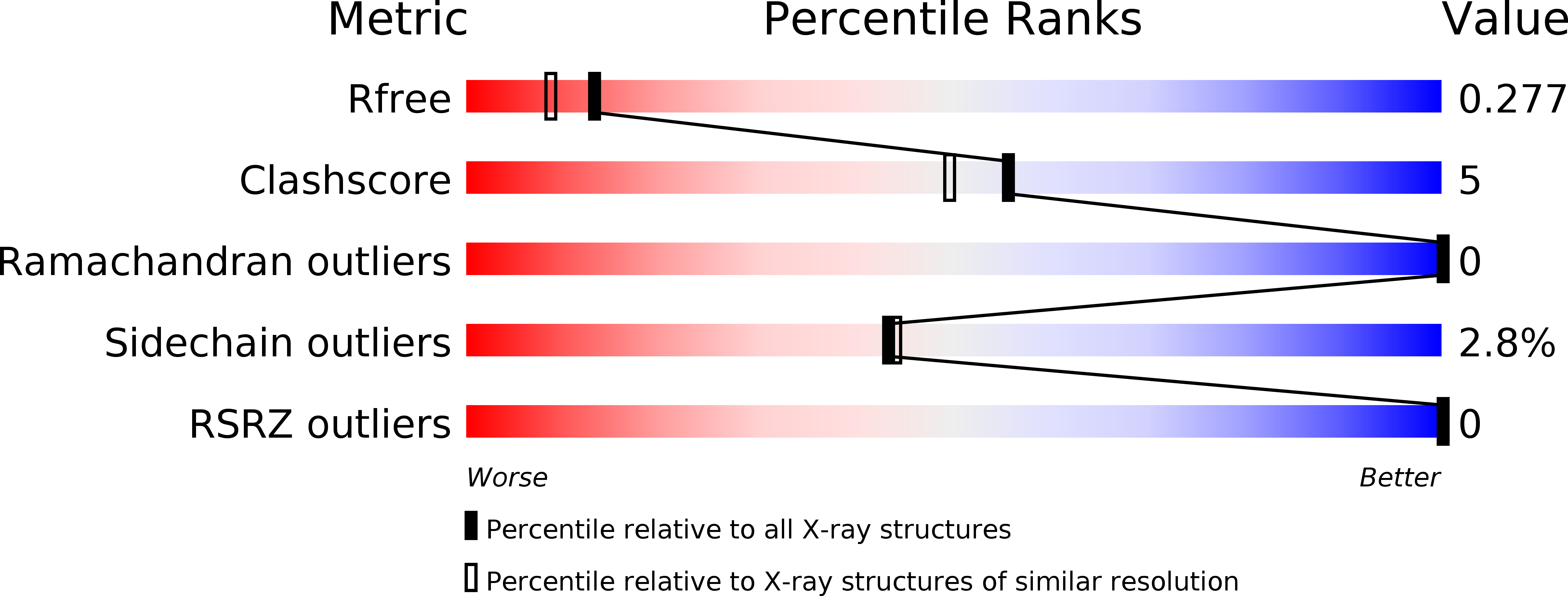
R-Value Free:
0.27
R-Value Work:
0.23
R-Value Observed:
0.23
Space Group:
P 1 21 1