
Deposition Date
2012-06-21
Release Date
2013-01-30
Last Version Date
2024-05-01
Entry Detail
PDB ID:
4AYQ
Keywords:
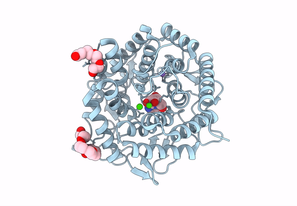
Title:
Structure of The GH47 processing alpha-1,2-mannosidase from Caulobacter strain K31 in complex with mannoimidazole
Biological Source:
Source Organism(s):
CAULOBACTER SP. (Taxon ID: 366602)
Expression System(s):
Method Details:
Experimental Method:
Resolution:
1.10 Å
R-Value Free:
0.10
R-Value Work:
0.08
R-Value Observed:
0.08
Space Group:
H 3
