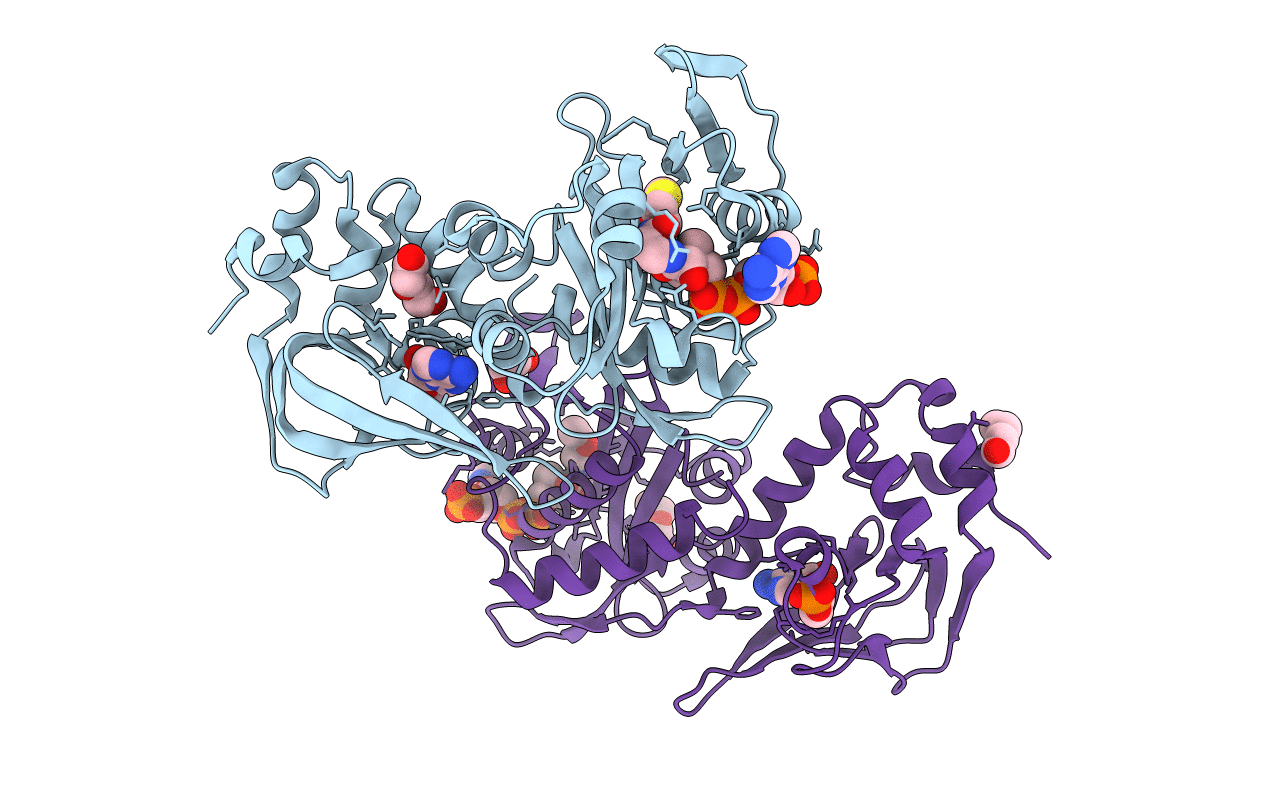
4AVC image
Entry Detail
PDB ID:
4AVC
Keywords:
Title:
Crystal structure of protein lysine acetyltransferase Rv0998 in complex with acetyl CoA and cAMP
Biological Source:
Host Organism:
PDB Version:
Deposition Date:
2012-05-24
Release Date:
2012-07-11
Method Details:
Experimental Method:
Resolution:
2.81 Å
R-Value Free:
0.26
R-Value Work:
0.19
R-Value Observed:
0.20
Space Group:
P 1 21 1
Macromolecular Entities
Polymer Type:polypeptide(L)
Description:LYSINE ACETYLTRANSFERASE
Chain IDs:A, B
Chain Length:333
Number of Molecules:2
Biological Source:MYCOBACTERIUM TUBERCULOSIS
Modified Residue
Compound ID Chain ID Parent Comp ID Details 2D Image
MSE A MET SELENOMETHIONINE
Primary Citation
Cyclic AMP Regulation of Protein Lysine Acetylation in Mycobacterium Tuberculosis.
Nat.Struct.Mol.Biol. 19 811 ? (2012)
PMID: 22773105 DOI: 10.1038/NSMB.2318

Abstact

Protein lysine acetylation networks can regulate central processes such as carbon metabolism and gene expression in bacteria. In Escherichia coli, cyclic AMP (cAMP) regulates protein lysine acetyltransferase (PAT) activity at the transcriptional level, but in Mycobacterium tuberculosis, fusion of a cyclic nucleotide-binding domain to a Gcn5-like PAT domain enables direct cAMP control of protein acetylation. Here we describe the allosteric activation mechanism of M. tuberculosis PAT. The crystal structures of the autoinhibited and cAMP-activated PAT reveal that cAMP binds to a cryptic site in the regulatory domain that is over 32 Å from the catalytic site. An extensive conformational rearrangement relieves this autoinhibition by means of a substrate-mimicking lid that covers the protein-substrate binding surface. A steric double latch couples the domains by harnessing a classic, cAMP-mediated conformational switch. The structures suggest general features that enable the evolution of long-range communication between linked domains.

Legend

Protein

Chemical

Disease

Primary Citation of related structures