4AUB image
Deposition Date 2012-05-16
Release Date 2012-06-06
Last Version Date 2023-12-20
Entry Detail
PDB ID:
4AUB
Keywords:
Title:
the complex Structure of the bacterial aldo-keto reductase AKR14A1 with NADP and citrate
Biological Source:
Source Organism:
Host Organism:
Method Details:
Experimental Method:
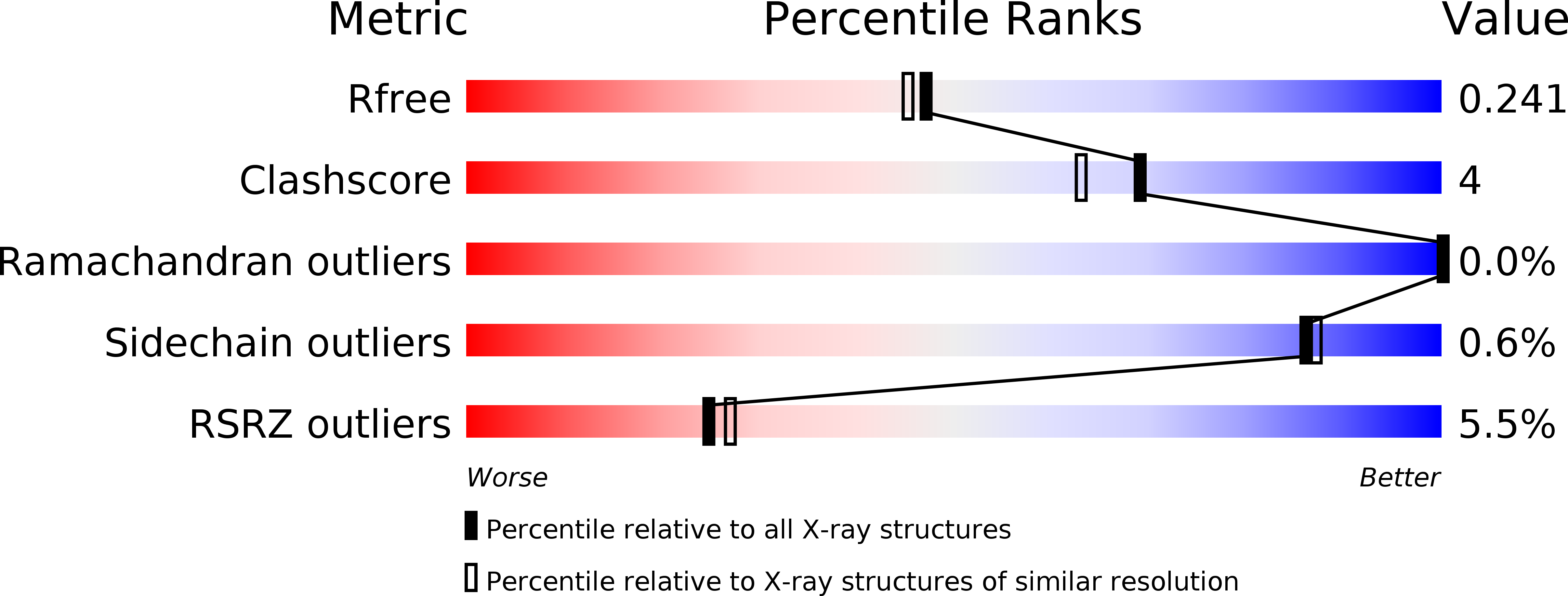
Resolution:
2.05 Å
R-Value Free:
0.23
R-Value Work:
0.20
R-Value Observed:
0.20
Space Group:
P 1 21 1
Macromolecular Entities
Polymer Type:polypeptide(L)
Molecule:ALDO-KETO REDUCTASE AKR14A1
Gene (Uniprot):gpr
Chain IDs:A, B, C, D, E, F, G, H
Chain Length:366
Number of Molecules:8
Biological Source:ESCHERICHIA COLI K-12
Primary Citation
The Diversity of Microbial Aldo/Keto Reductases from Escherichia Coli K12.
Chem.Biol.Interact 202 168 ? (2013)
PMID: 23103600 DOI: 10.1016/J.CBI.2012.10.008

Abstact

The genome of Escherichia coli K12 contains 9 open reading frames encoding aldo/keto reductases (AKRs) that are differentially regulated and sequence diverse. A significant amount of data is available for the E. coli AKRs through the availability of gene knockouts and gene expression studies, which adds to the biochemical and kinetic data. This together with the availability of crystal structures for nearly half of the E. coli AKRs and homologues of several others provides an opportunity to look at the diversity of these representative bacterial AKRs. Based around the common AKR fold of (β/α)8 barrel with two additional α-helices, the E. coli AKRs have a loop structure that is more diverse than their mammalian counterparts, creating a variety of active site architectures. Nearly half of the AKRs are expected to be monomeric, but there are examples of dimeric, trimeric and octameric enzymes, as well as diversity in specificity for NAD as well as NADP as a cofactor. However in functional assignments and characterisation of enzyme activities there is a paucity of data when compared to the mammalian AKR enzymes.

Legend

Protein

Chemical

Disease

Primary Citation of related structures