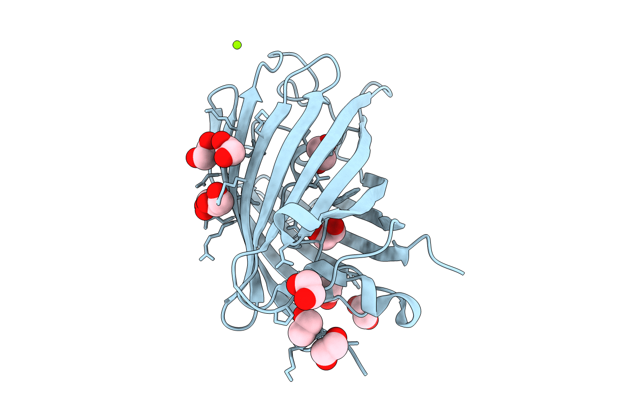
4AS8 image
Deposition Date 2012-04-29
Release Date 2012-10-31
Last Version Date 2024-11-13
Entry Detail
PDB ID:
4AS8
Title:
X-ray structure of the cyan fluorescent protein Cerulean cryoprotected with ethylene glycol
Biological Source:
Source Organism:
Host Organism:
Method Details:
Experimental Method:
Resolution:
1.02 Å
R-Value Free:
0.13
R-Value Work:
0.11
R-Value Observed:
0.11
Space Group:
P 21 21 21
Macromolecular Entities
Polymer Type:polypeptide(L)
Molecule:GREEN FLUORESCENT PROTEIN
Gene (Uniprot):GFP
Mutations:YES
Chain IDs:A
Chain Length:245
Number of Molecules:1
Biological Source:AEQUOREA VICTORIA
Modified Residue
Compound ID Chain ID Parent Comp ID Details 2D Image
CRF A THR ?
Primary Citation
Alteration of Fluorescent Protein Spectroscopic Properties Upon Cryoprotection
Acta Crystallogr.,Sect.D 68 1578 ? (2012)
PMID: 23090407 DOI: 10.1107/S0907444912037900

Abstact

Cryoprotection of a protein crystal by addition of small-molecule compounds may sometimes affect the structure of its active site. The spectroscopic and structural effects of the two cryoprotectants glycerol and ethylene glycol on the cyan fluorescent protein Cerulean were investigated. While glycerol had almost no noticeable effect, ethylene glycol was shown to induce a systematic red shift of the UV-vis absorption and fluorescence emission spectra. Additionally, ethylene glycol molecules were shown to enter the core of the protein, with one of them binding in close vicinity to the chromophore, which provides a sound explanation for the observed spectroscopic changes. These results highlight the need to systematically record spectroscopic data on crystals of light-absorbing proteins and reinforce the notion that fluorescent proteins must not been seen as rigid structures.

Legend

Protein

Chemical

Disease