4AQR image
Deposition Date 2012-04-19
Release Date 2012-10-17
Last Version Date 2024-05-08
Entry Detail
PDB ID:
4AQR
Title:
Crystal structure of calmodulin in complex with the regulatory domain of a plasma-membrane Ca2+-ATPase
Biological Source:
Source Organism:
Host Organism:
Method Details:
Experimental Method:
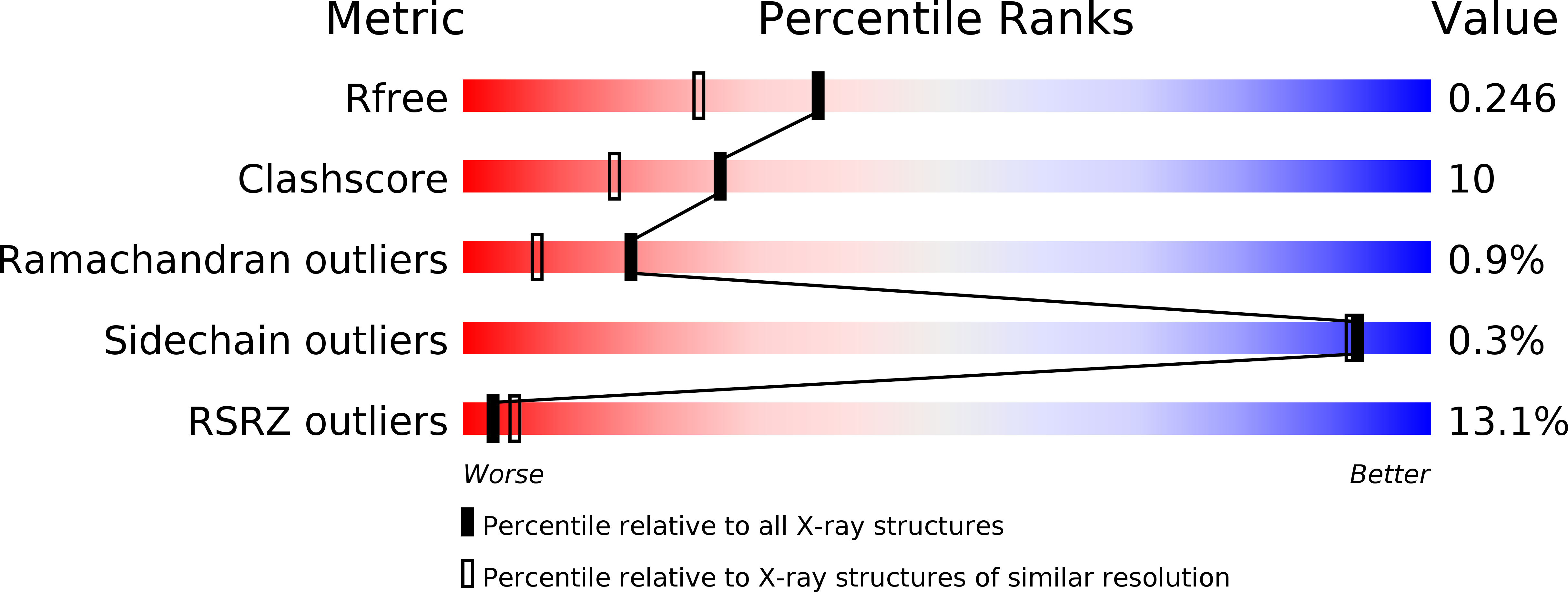
Resolution:
1.95 Å
R-Value Free:
0.24
R-Value Work:
0.21
R-Value Observed:
0.21
Space Group:
P 41 21 2
Macromolecular Entities
Structures with similar UniProt ID
Protein Blast
Polymer Type:polypeptide(L)
Molecule:CALMODULIN-7
Gene (Uniprot):CAM7
Chain IDs:A, B
Chain Length:149
Number of Molecules:2
Biological Source:ARABIDOPSIS THALIANA
Structures with similar UniProt ID
Protein Blast
Polymer Type:polypeptide(L)
Molecule:CALCIUM-TRANSPORTING ATPASE 8, PLASMA MEMBRANE-TYPE
Gene (Uniprot):ACA8
Chain IDs:C (auth: D)
Chain Length:57
Number of Molecules:1
Biological Source:ARABIDOPSIS THALIANA
Ligand Molecules
Primary Citation
A Bimodular Mechanism of Calcium Control in Eukaryotes
Nature 491 468 ? (2012)
PMID: 23086147 DOI: 10.1038/NATURE11539

Abstact

Calcium ions (Ca(2+)) have an important role as secondary messengers in numerous signal transduction processes, and cells invest much energy in controlling and maintaining a steep gradient between intracellular (∼0.1-micromolar) and extracellular (∼2-millimolar) Ca(2+) concentrations. Calmodulin-stimulated calcium pumps, which include the plasma-membrane Ca(2+)-ATPases (PMCAs), are key regulators of intracellular Ca(2+) in eukaryotes. They contain a unique amino- or carboxy-terminal regulatory domain responsible for autoinhibition, and binding of calcium-loaded calmodulin to this domain releases autoinhibition and activates the pump. However, the structural basis for the activation mechanism is unknown and a key remaining question is how calmodulin-mediated PMCA regulation can cover both basal Ca(2+) levels in the nanomolar range as well as micromolar-range Ca(2+) transients generated by cell stimulation. Here we present an integrated study combining the determination of the high-resolution crystal structure of a PMCA regulatory-domain/calmodulin complex with in vivo characterization and biochemical, biophysical and bioinformatics data that provide mechanistic insights into a two-step PMCA activation mechanism mediated by calcium-loaded calmodulin. The structure shows the entire PMCA regulatory domain and reveals an unexpected 2:1 stoichiometry with two calcium-loaded calmodulin molecules binding to different sites on a long helix. A multifaceted characterization of the role of both sites leads to a general structural model for calmodulin-mediated regulation of PMCAs that allows stringent, highly responsive control of intracellular calcium in eukaryotes, making it possible to maintain a stable, basal level at a threshold Ca(2+) concentration, where steep activation occurs.

Legend

Protein

Chemical

Disease

Primary Citation of related structures