4AP5 image
Deposition Date 2012-03-30
Release Date 2012-08-08
Last Version Date 2024-11-06
Entry Detail
PDB ID:
4AP5
Keywords:
Title:
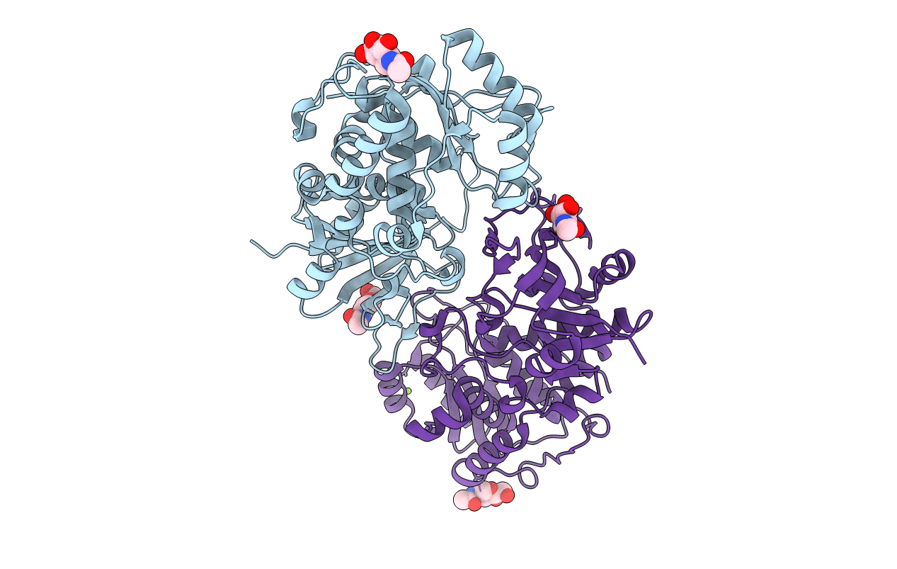
Crystal structure of human POFUT2
Biological Source:
Source Organism:
HOMO SAPIENS (Taxon ID: 9606)
Host Organism:
Method Details:
Experimental Method:
Resolution:
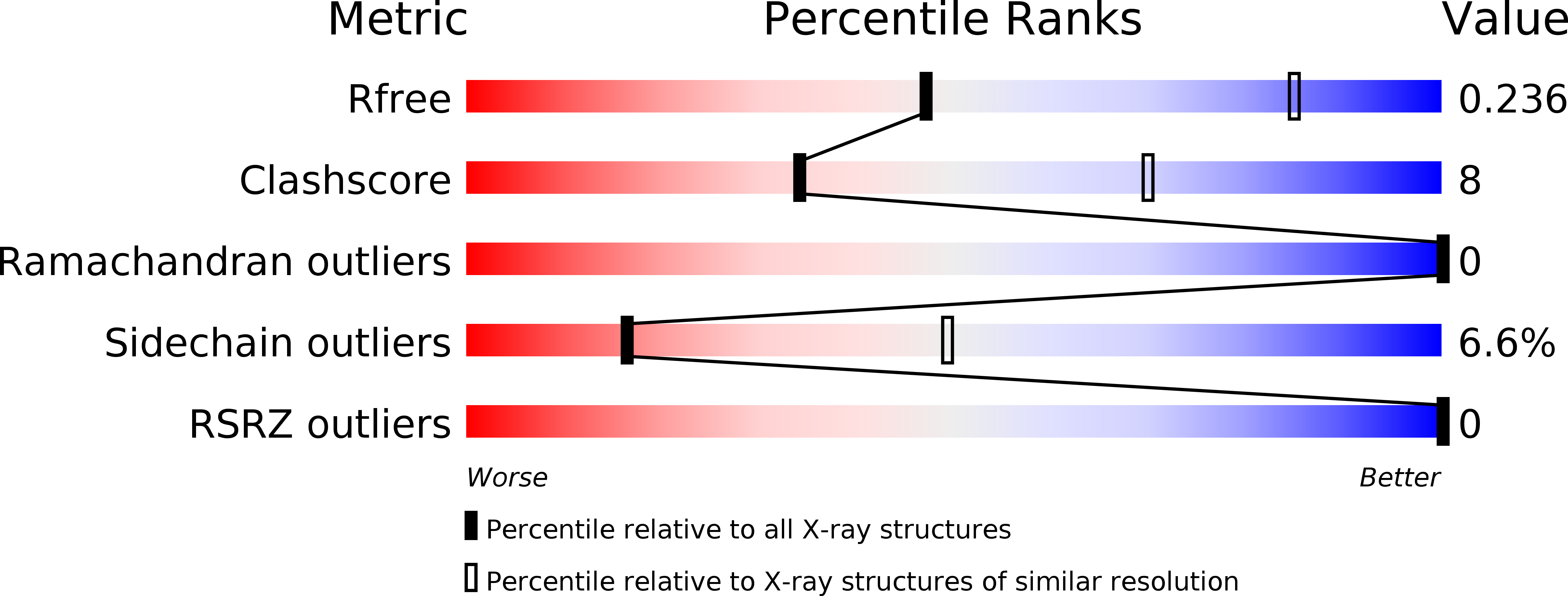
3.00 Å
R-Value Free:
0.23
R-Value Work:
0.17
R-Value Observed:
0.17
Space Group:
P 32 2 1
Macromolecular Entities
Structures with similar UniProt ID
Protein Blast
Polymer Type:polypeptide(L)
Molecule:GDP-FUCOSE PROTEIN O-FUCOSYLTRANSFERASE 2
Gene (Uniprot):POFUT2
Chain IDs:A, B
Chain Length:408
Number of Molecules:2
Biological Source:HOMO SAPIENS
Modified Residue
Compound ID Chain ID Parent Comp ID Details 2D Image
ASN A ASN GLYCOSYLATION SITE
Primary Citation
Structure of Human Pofut2: Insights Into Thrombospondin Type 1 Repeat Fold and O-Fucosylation.
Embo J. 31 3183 ? (2012)
PMID: 22588082 DOI: 10.1038/EMBOJ.2012.143

Abstact

Protein O-fucosylation is a post-translational modification found on serine/threonine residues of thrombospondin type 1 repeats (TSR). The fucose transfer is catalysed by the protein O-fucosyltransferase 2 (POFUT2) and >40 human proteins contain the TSR consensus sequence for POFUT2-dependent fucosylation. To better understand O-fucosylation on TSR, we carried out a structural and functional analysis of human POFUT2 and its TSR substrate. Crystal structures of POFUT2 reveal a variation of the classical GT-B fold and identify sugar donor and TSR acceptor binding sites. Structural findings are correlated with steady-state kinetic measurements of wild-type and mutant POFUT2 and TSR and give insight into the catalytic mechanism and substrate specificity. By using an artificial mini-TSR substrate, we show that specificity is not primarily encoded in the TSR protein sequence but rather in the unusual 3D structure of a small part of the TSR. Our findings uncover that recognition of distinct conserved 3D fold motifs can be used as a mechanism to achieve substrate specificity by enzymes modifying completely folded proteins of very wide sequence diversity and biological function.

Legend

Protein

Chemical

Disease

Primary Citation of related structures