4AP4 image
Entry Detail
PDB ID:
4AP4
Title:
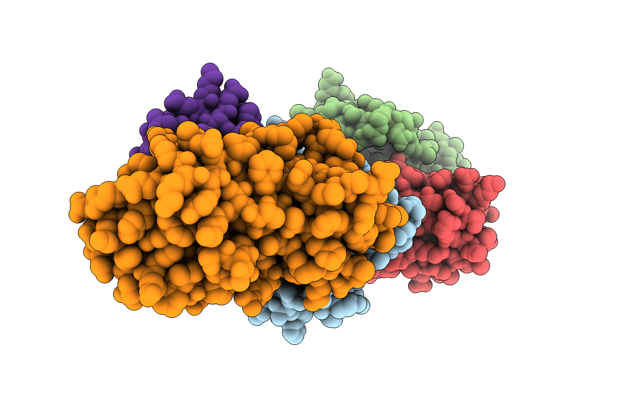
Rnf4 - ubch5a - ubiquitin heterotrimeric complex
Biological Source:
Host Organism:
PDB Version:
Deposition Date:
2012-03-30
Release Date:
2012-07-25
Method Details:
Experimental Method:
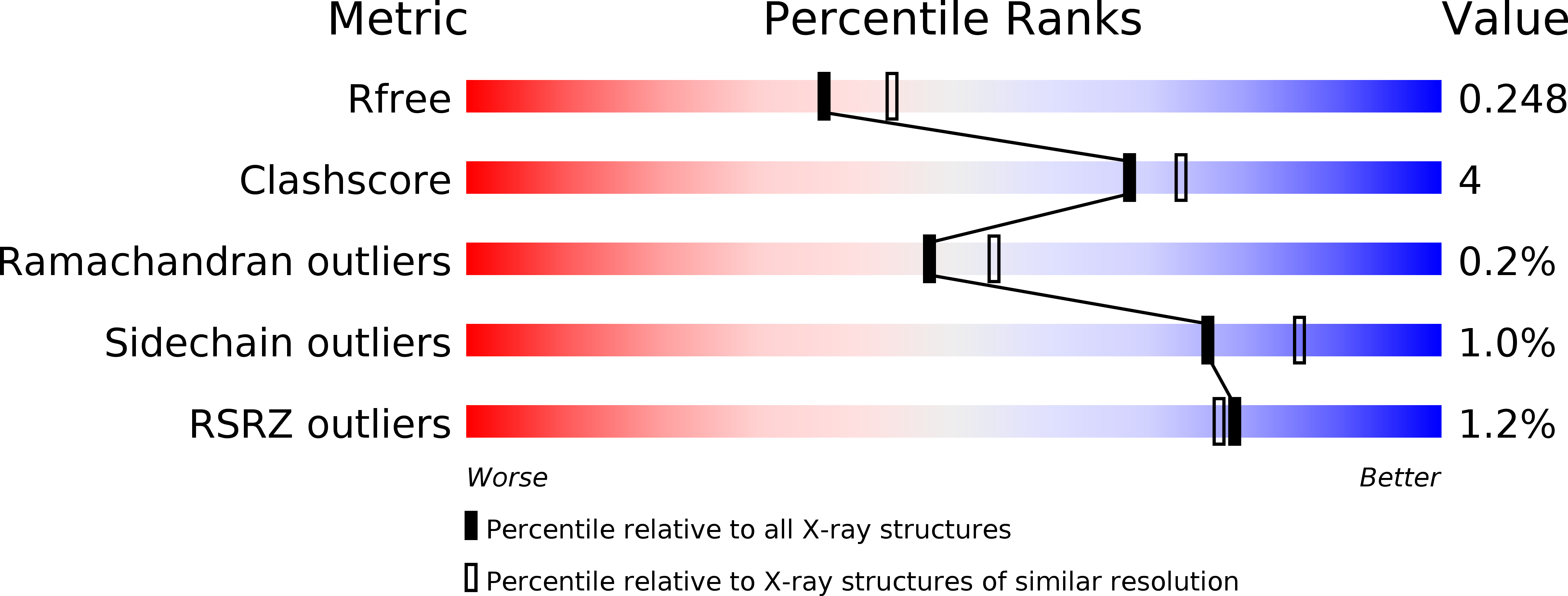
Resolution:
2.21 Å
R-Value Free:
0.24
R-Value Work:
0.20
R-Value Observed:
0.20
Space Group:
P 21 21 21
Macromolecular Entities
Polymer Type:polypeptide(L)
Description:E3 UBIQUITIN LIGASE RNF4
Chain IDs:A
Chain Length:133
Number of Molecules:1
Biological Source:RATTUS NORVEGICUS
Polymer Type:polypeptide(L)
Description:UBIQUITIN-CONJUGATING ENZYME E2 D1
Mutations:YES
Chain IDs:B, D (auth: E)
Chain Length:153
Number of Molecules:2
Biological Source:HOMO SAPIENS
Polymer Type:polypeptide(L)
Description:UBIQUITIN C
Chain IDs:C, E (auth: F)
Chain Length:80
Number of Molecules:2
Biological Source:HOMO SAPIENS
Ligand Molecules
Primary Citation
Structure of a Ring E3 Ligase and Ubiquitin-Loaded E2 Primed for Catalysis
Nature 489 115 ? (2012)
PMID: 22842904 DOI: 10.1038/NATURE11376

Abstact

Ubiquitin modification is mediated by a large family of specificity determining ubiquitin E3 ligases. To facilitate ubiquitin transfer, RING E3 ligases bind both substrate and a ubiquitin E2 conjugating enzyme linked to ubiquitin via a thioester bond, but the mechanism of transfer has remained elusive. Here we report the crystal structure of the dimeric RING domain of rat RNF4 in complex with E2 (UbcH5A) linked by an isopeptide bond to ubiquitin. While the E2 contacts a single protomer of the RING, ubiquitin is folded back onto the E2 by contacts from both RING protomers. The carboxy-terminal tail of ubiquitin is locked into an active site groove on the E2 by an intricate network of interactions, resulting in changes at the E2 active site. This arrangement is primed for catalysis as it can deprotonate the incoming substrate lysine residue and stabilize the consequent tetrahedral transition-state intermediate.

Legend

Protein

Chemical

Disease

Primary Citation of related structures