4ANE image
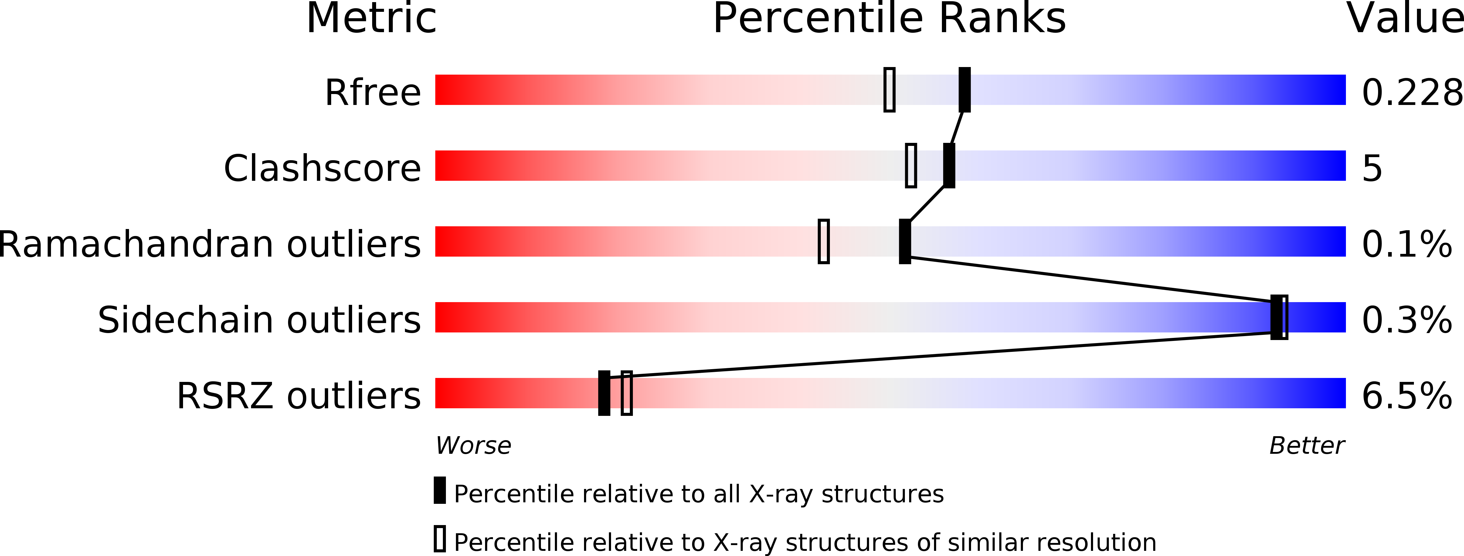
Deposition Date 2012-03-16
Release Date 2013-03-13
Last Version Date 2023-12-20
Entry Detail
PDB ID:
4ANE
Keywords:
Title:
R80N MUTANT OF NUCLEOSIDE DIPHOSPHATE KINASE FROM MYCOBACTERIUM TUBERCULOSIS
Biological Source:
Source Organism:
Host Organism:
Method Details:
Experimental Method:
Resolution:
1.90 Å
R-Value Free:
0.23
R-Value Work:
0.18
R-Value Observed:
0.18
Space Group:
I 1 2 1
Macromolecular Entities
Structures with similar UniProt ID
Protein Blast
Polymer Type:polypeptide(L)
Molecule:NUCLEOSIDE DIPHOSPHATE KINASE
Mutagens:YES
Chain IDs:A, B, C, D, E, F
Chain Length:136
Number of Molecules:6
Biological Source:MYCOBACTERIUM TUBERCULOSIS
Ligand Molecules
Primary Citation
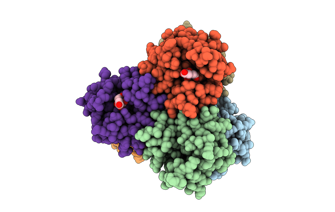
Structure of Mycobacterium Tuberculosis Nucleoside Diphosphate Kinase R80N Mutant in Complex with Citrate
Acta Crystallogr.,Sect.D 70 40 ? (2014)
PMID: 24419614 DOI: 10.1107/S2053230X13034134

Abstact

The crystal structure of the wild-type nucleoside diphosphate kinase from Mycobacterium tuberculosis at 2.6 Å resolution revealed that the intersubunit salt bridge Arg80-Asp93 contributes to the thermal stability of the hexamer (Tm = 76°C). On mutating Asp93 to Asn to break the salt bridge, the thermal stability dramatically decreased by 27.6°C. Here, on mutating Arg80 to Asn, the thermal stability also significantly decreased by 8.0°C. In the X-ray structure of the R80N mutant solved at 1.9 Å resolution the salt bridge was replaced by intersubunit hydrogen bonds that contribute to the thermal stability of the hexamer. A citrate anion from the crystallization buffer was bound at the bottom of the nucleotide-binding site via electrostatic and hydrogen-bonding interactions with six conserved residues involved in nucleotide binding. Structural analysis shows that the citrate is present at the location of the nucleotide phosphate groups.

Legend

Protein

Chemical

Disease

Primary Citation of related structures