4AM3 image
Deposition Date 2012-03-07
Release Date 2012-04-18
Last Version Date 2023-12-20
Entry Detail
PDB ID:
4AM3
Keywords:
Title:
Crystal structure of C. crescentus PNPase bound to RNA
Biological Source:
Source Organism:
Host Organism:
Method Details:
Experimental Method:
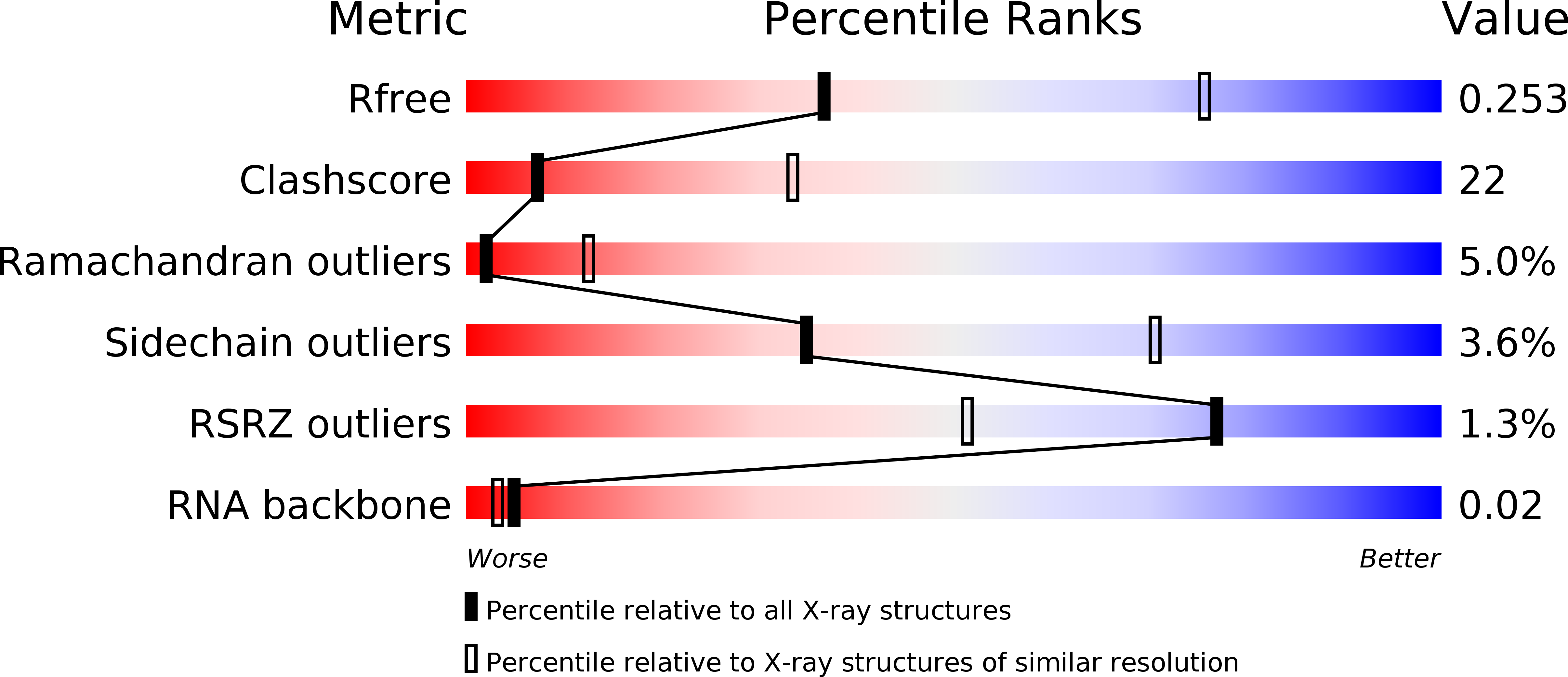
Resolution:
3.00 Å
R-Value Free:
0.25
R-Value Work:
0.20
R-Value Observed:
0.21
Space Group:
P 2 21 21
Macromolecular Entities
Structures with similar UniProt ID
Protein Blast
Polymer Type:polypeptide(L)
Molecule:POLYRIBONUCLEOTIDE NUCLEOTIDYLTRANSFERASE
Gene (Uniprot):pnp
Chain IDs:A, B, C
Chain Length:717
Number of Molecules:3
Biological Source:CAULOBACTER VIBRIOIDES
Polymer Type:polyribonucleotide
Molecule:RNA, 5'-R(*UP*AP*AP*CP*UP*UP*UP*GP*GP)-3'
Chain IDs:D, E, F (auth: H), G (auth: I)
Chain Length:9
Number of Molecules:4
Biological Source:ESCHERICHIA COLI
Ligand Molecules
Primary Citation
Crystal Structure of Caulobacter Crescentus Polynucleotide Phosphorylase Reveals a Mechanism of RNA Substrate Channelling and RNA Degradosome Assembly.
Open Biol. 2 20028 ? (2012)
PMID: 22724061 DOI: 10.1098/RSOB.120028

Abstact

Polynucleotide phosphorylase (PNPase) is an exoribonuclease that cleaves single-stranded RNA substrates with 3'-5' directionality and processive behaviour. Its ring-like, trimeric architecture creates a central channel where phosphorolytic active sites reside. One face of the ring is decorated with RNA-binding K-homology (KH) and S1 domains, but exactly how these domains help to direct the 3' end of single-stranded RNA substrates towards the active sites is an unsolved puzzle. Insight into this process is provided by our crystal structures of RNA-bound and apo Caulobacter crescentus PNPase. In the RNA-free form, the S1 domains adopt a 'splayed' conformation that may facilitate capture of RNA substrates. In the RNA-bound structure, the three KH domains collectively close upon the RNA and direct the 3' end towards a constricted aperture at the entrance of the central channel. The KH domains make non-equivalent interactions with the RNA, and there is a marked asymmetry within the catalytic core of the enzyme. On the basis of these data, we propose that structural non-equivalence, induced upon RNA binding, helps to channel substrate to the active sites through mechanical ratcheting. Structural and biochemical analyses also reveal the basis for PNPase association with RNase E in the multi-enzyme RNA degradosome assembly of the α-proteobacteria.

Legend

Protein

Chemical

Disease

Primary Citation of related structures