Deposition Date
2012-02-24
Release Date
2012-08-15
Last Version Date
2024-10-09
Entry Detail
PDB ID:
4AKM
Keywords:
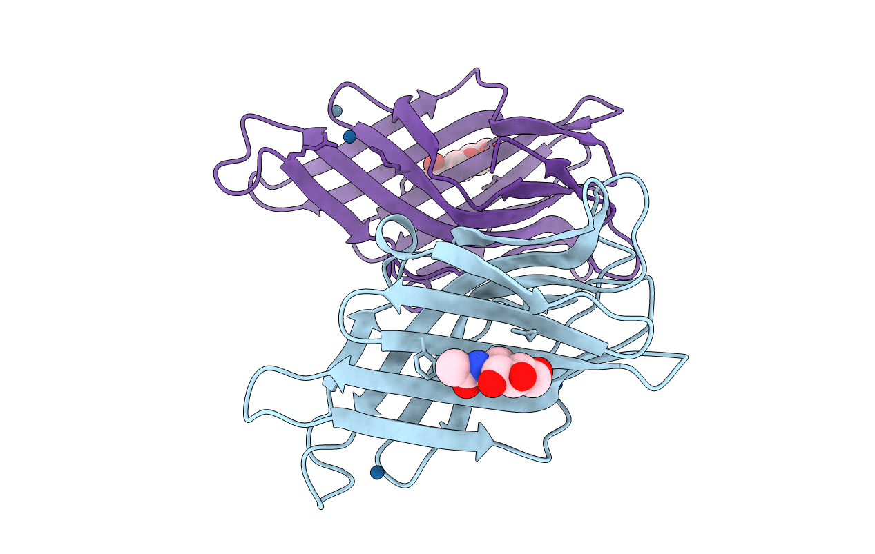
Title:
Crystal structure of the human lysosome-associated membrane protein LAMP-3 (aka DC-LAMP)
Biological Source:
Source Organism(s):
HOMO SAPIENS (Taxon ID: 9606)
Expression System(s):
Method Details:
Experimental Method:
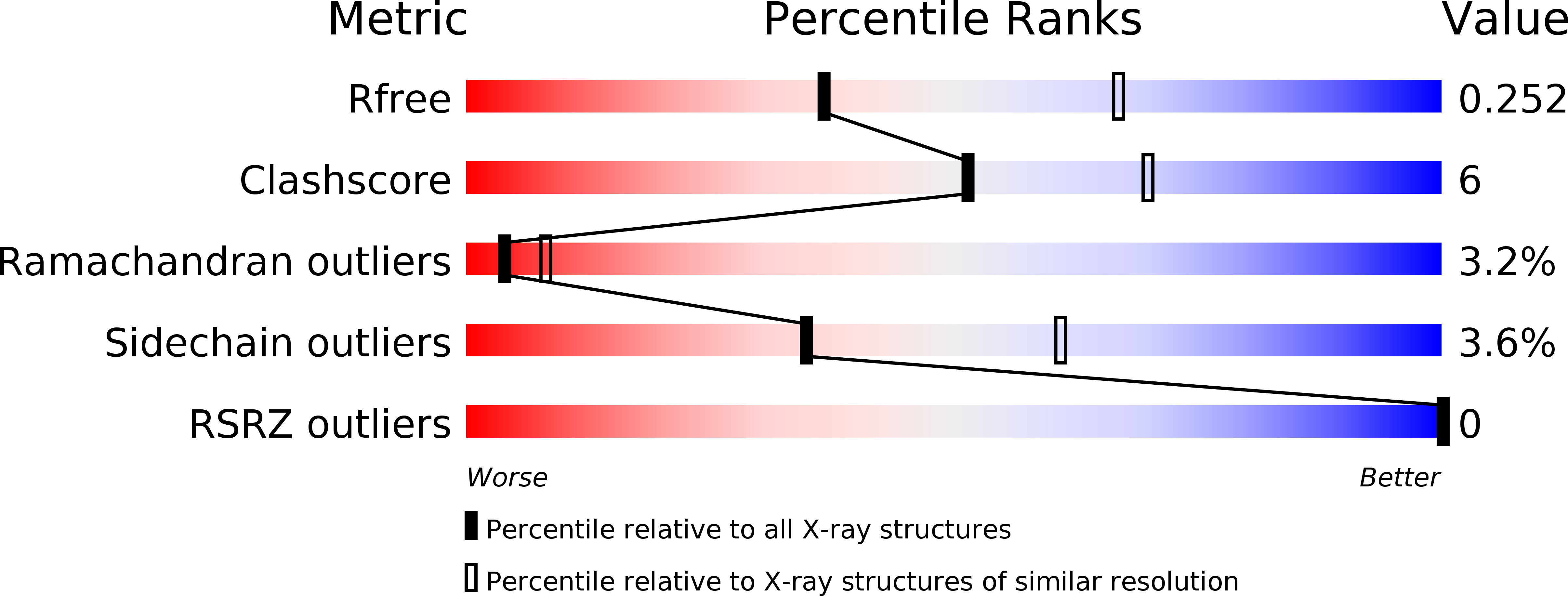
Resolution:
2.69 Å
R-Value Free:
0.24
R-Value Work:
0.22
R-Value Observed:
0.22
Space Group:
P 31