4AK4 image
Entry Detail
PDB ID:
4AK4
Title:
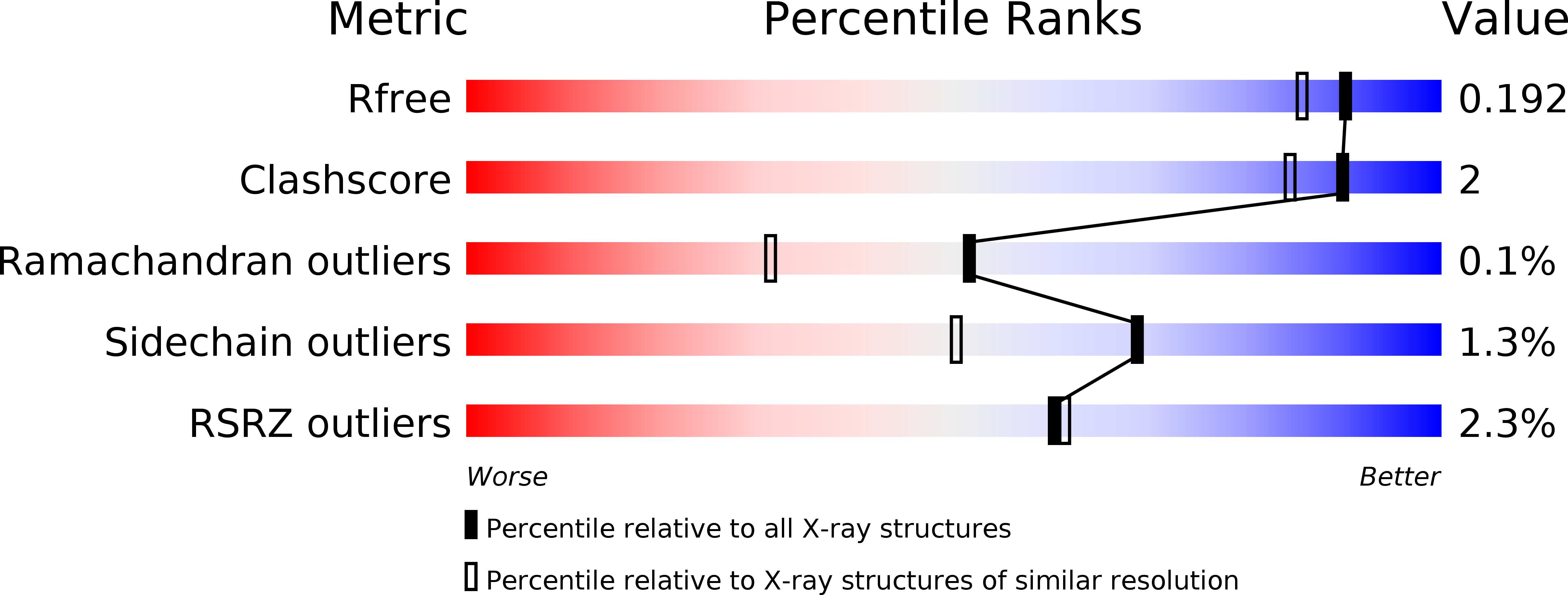
High resolution structure of Galactose Binding lectin from Champedak (CGB)
Biological Source:
Source Organism:
PDB Version:
Deposition Date:
2012-02-21
Release Date:
2013-02-27
Method Details:
Experimental Method:
Resolution:
1.65 Å
R-Value Free:
0.18
R-Value Work:
0.16
R-Value Observed:
0.16
Space Group:
P 1 21 1
Macromolecular Entities
Polymer Type:polypeptide(L)
Description:AGGLUTININ ALPHA CHAIN
Chain IDs:A, C, E, G, I, K, M, O
Chain Length:133
Number of Molecules:8
Biological Source:ARTOCARPUS INTEGER
Polymer Type:polypeptide(L)
Description:AGGLUTININ BETA-4 CHAIN
Chain IDs:B, D, F, H, J, L, N, P
Chain Length:21
Number of Molecules:8
Biological Source:ARTOCARPUS INTEGER
Ligand Molecules
Primary Citation
Structures and Binding Specificity of Galactose- and Mannose-Binding Lectins from Champedak: Differences from Jackfruit Lectins
Acta Crystallogr.,Sect.F 70 709 ? (2014)
PMID: 24915077 DOI: 10.1107/S2053230X14008966

Abstact

Galactose-binding and mannose-binding lectins from the champedak fruit, which is native to South-east Asia, exhibit useful potential clinical applications. The specificity of the two lectins for their respective ligands allows the detection of potential cancer biomarkers and monitoring of the glycosylated state of proteins in human serum and/or urine. To fully understand and expand the use of these natural proteins, their complete sequences and crystal structures are presented here, together with details of sugar binding.

Legend

Protein

Chemical

Disease

Primary Citation of related structures