4AJC image
Entry Detail
PDB ID:
4AJC
Keywords:
Title:
3D structure of E. coli Isocitrate Dehydrogenase K100M mutant in complex with alpha-ketoglutarate, calcium(II) and adenine nucleotide phosphate
Biological Source:
Source Organism:
Host Organism:
PDB Version:
Deposition Date:
2012-02-16
Release Date:
2012-10-31
Method Details:
Experimental Method:
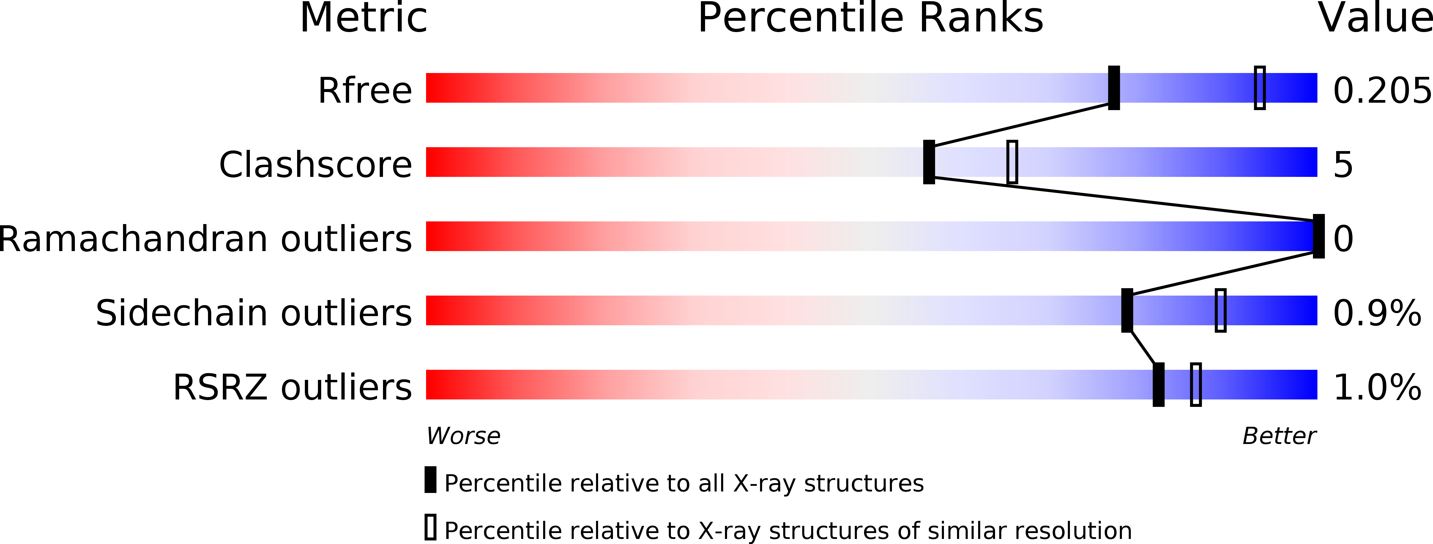
Resolution:
2.30 Å
R-Value Free:
0.20
R-Value Work:
0.17
R-Value Observed:
0.17
Space Group:
P 43 21 2
Macromolecular Entities
Polymer Type:polypeptide(L)
Description:NADP ISOCITRATE DEHYDROGENASE
Mutations:YES
Chain IDs:A
Chain Length:416
Number of Molecules:1
Biological Source:ESCHERICHIA COLI
Primary Citation
Induced Fit and the Catalytic Mechanism of Isocitrate Dehydrogenase.
Biochemistry 51 7098 ? (2012)
PMID: 22891681 DOI: 10.1021/BI300483W

Abstact

NADP(+) dependent isocitrate dehydrogenase (IDH; EC 1.1.1.42) belongs to a large family of α-hydroxyacid oxidative β-decarboxylases that catalyze similar three-step reactions, with dehydrogenation to an oxaloacid intermediate preceding β-decarboxylation to an enol intermediate followed by tautomerization to the final α-ketone product. A comprehensive view of the induced fit needed for catalysis is revealed on comparing the first "fully closed" crystal structures of a pseudo-Michaelis complex of wild-type Escherichia coli IDH (EcoIDH) and the "fully closed" reaction product complex of the K100M mutant with previously obtained "quasi-closed" and "open" conformations. Conserved catalytic residues, binding the nicotinamide ring of NADP(+) and the metal-bound substrate, move as rigid bodies during domain closure by a hinge motion that spans the central β-sheet in each monomer. Interactions established between Thr105 and Ser113, which flank the "phosphorylation loop", and the nicotinamide mononucleotide moiety of NADP(+) establish productive coenzyme binding. Electrostatic interactions of a Lys100-Leu103-Asn115-Glu336 tetrad play a pivotal role in assembling a catalytically competent active site. As predicted, Lys230* is positioned to deprotonate/reprotonate the α-hydroxyl in both reaction steps and Tyr160 moves into position to protonate C3 following β-decarboxylation. A proton relay from the catalytic triad Tyr160-Asp307-Lys230* connects the α-hydroxyl of isocitrate to the bulk solvent to complete the picture of the catalytic mechanism.

Legend

Protein

Chemical

Disease

Primary Citation of related structures