4AIR image
Deposition Date 2012-02-13
Release Date 2012-02-29
Last Version Date 2024-11-13
Entry Detail
PDB ID:
4AIR
Keywords:
Title:
Leishmania major cysteine synthase
Biological Source:
Source Organism:
Host Organism:
Method Details:
Experimental Method:
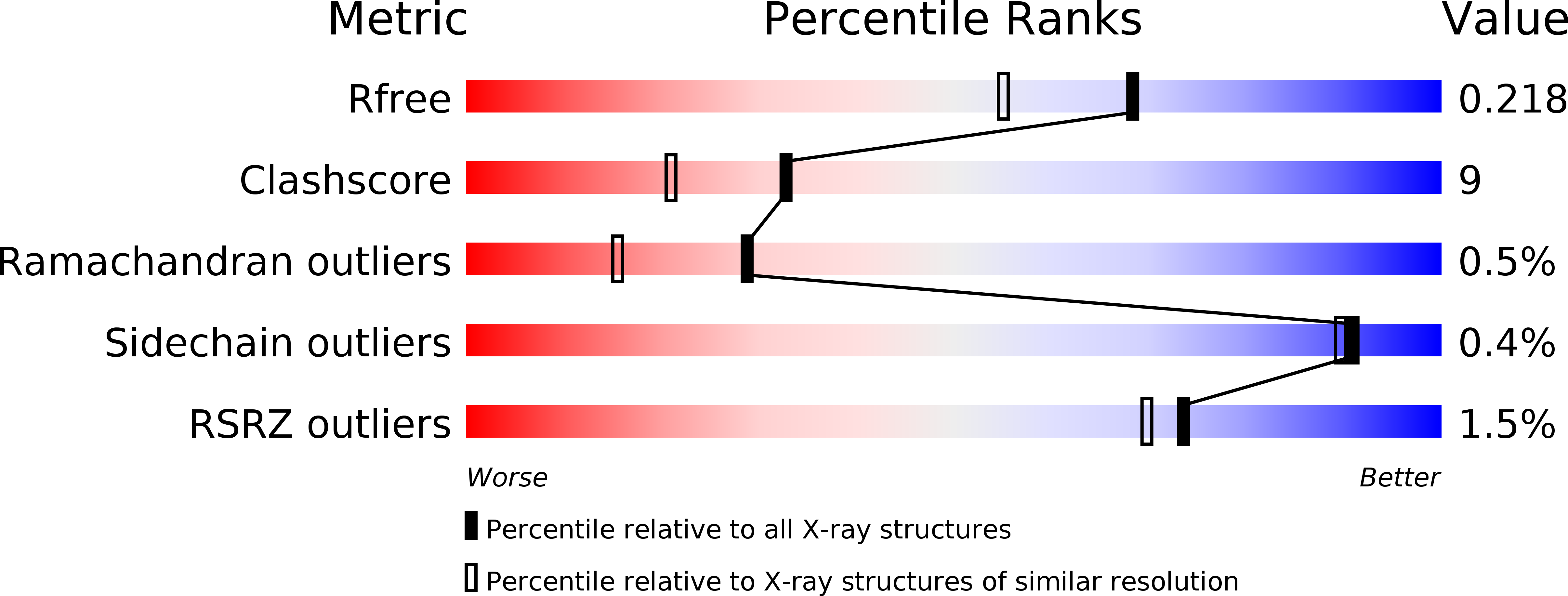
Resolution:
1.80 Å
R-Value Free:
0.20
R-Value Work:
0.15
R-Value Observed:
0.15
Space Group:
P 21 21 21
Macromolecular Entities
Structures with similar UniProt ID
Protein Blast
Polymer Type:polypeptide(L)
Molecule:Cysteine synthase
Gene (Uniprot):LMJF_36_3590
Chain IDs:A, B
Chain Length:354
Number of Molecules:2
Biological Source:Leishmania major
Polymer Type:polypeptide(L)
Molecule:FGA-FGA-FGA-FGA-FGA
Chain IDs:C
Chain Length:5
Number of Molecules:1
Biological Source:Synthetic construct
Polymer Type:polypeptide(L)
Molecule:FGA-FGA-FGA-FGA
Chain IDs:D
Chain Length:4
Number of Molecules:1
Biological Source:Synthetic construct
Ligand Molecules
Primary Citation
Structure of Leishmania Major Cysteine Synthase.
Acta Crystallogr.,Sect.F 68 738 ? (2012)
PMID: 22750854 DOI: 10.1107/S1744309112019124

Abstact

Cysteine biosynthesis is a potential target for drug development against parasitic Leishmania species; these protozoa are responsible for a range of serious diseases. To improve understanding of this aspect of Leishmania biology, a crystallographic and biochemical study of L. major cysteine synthase has been undertaken, seeking to understand its structure, enzyme activity and modes of inhibition. Active enzyme was purified, assayed and crystallized in an orthorhombic form with a dimer in the asymmetric unit. Diffraction data extending to 1.8 Å resolution were measured and the structure was solved by molecular replacement. A fragment of γ-poly-D-glutamic acid, a constituent of the crystallization mixture, was bound in the enzyme active site. Although a D-glutamate tetrapeptide had insignificant inhibitory activity, the enzyme was competitively inhibited (K(i) = 4 µM) by DYVI, a peptide based on the C-terminus of the partner serine acetyltransferase with which the enzyme forms a complex. The structure surprisingly revealed that the cofactor pyridoxal phosphate had been lost during crystallization.

Legend

Protein

Chemical

Disease

Primary Citation of related structures