
Deposition Date
2012-02-08
Release Date
2012-02-15
Last Version Date
2023-12-20
Entry Detail
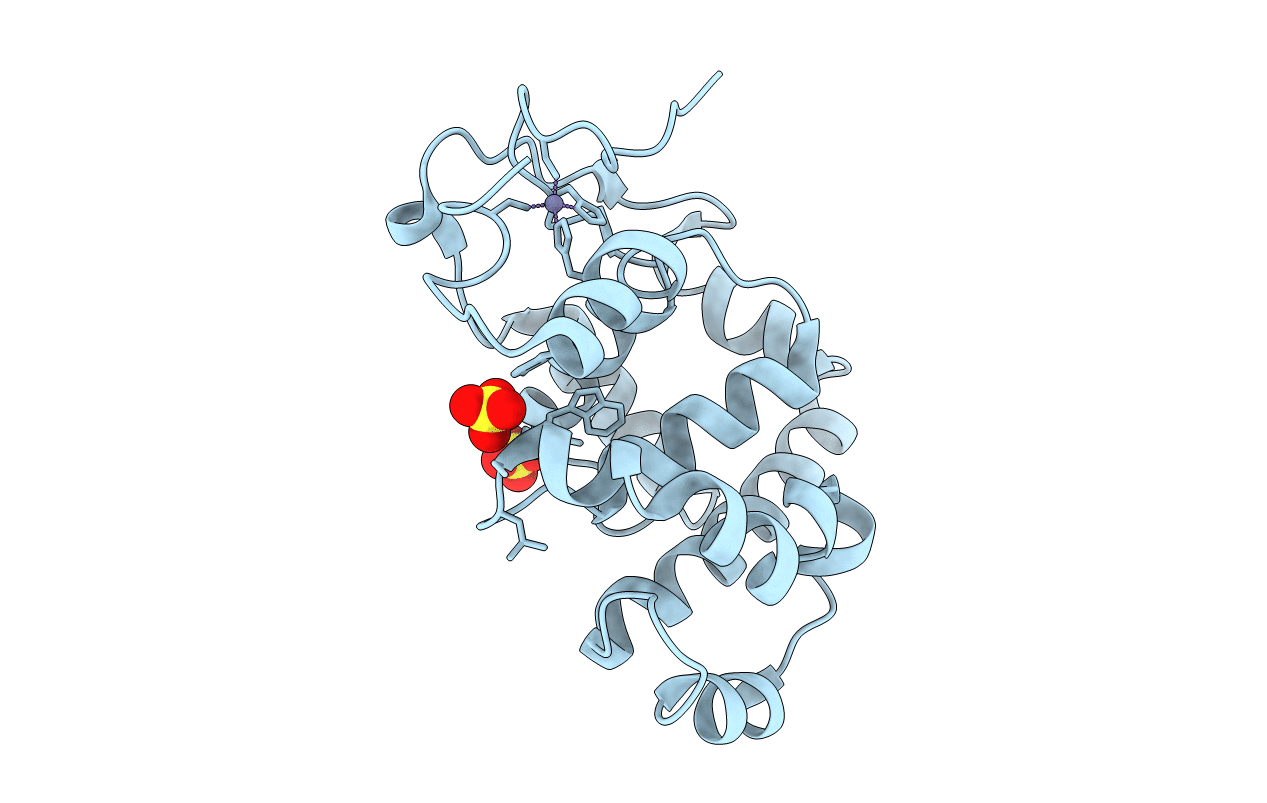
PDB ID:
4AI4
Keywords:
Title:
crystal structure of E38Q mutant of 3-methyladenine DNA glycosylase I from Staphylococcus aureus
Biological Source:
Source Organism(s):
STAPHYLOCOCCUS AUREUS SUBSP. AUREUS MSSA476 (Taxon ID: 282459)
Expression System(s):
Method Details:
Experimental Method:
Resolution:
1.73 Å
R-Value Free:
0.20
R-Value Work:
0.17
R-Value Observed:
0.17
Space Group:
C 1 2 1
