Deposition Date
2012-01-26
Release Date
2012-08-15
Last Version Date
2023-12-20
Entry Detail
PDB ID:
4AGE
Keywords:
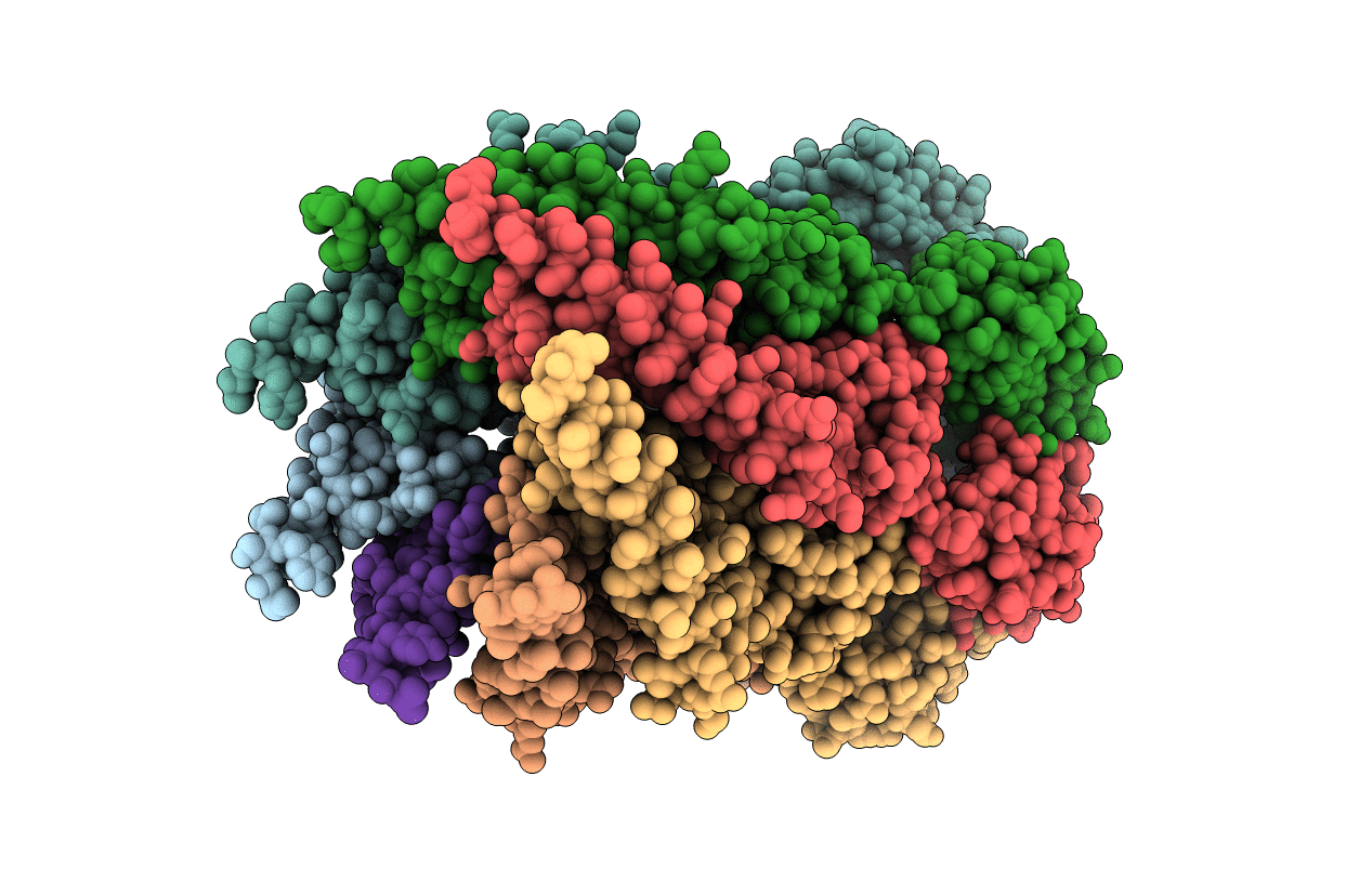
Title:
MTSSL spin labeled D67C mutant of MscS in the open form
Biological Source:
Source Organism(s):
ESCHERICHIA COLI (Taxon ID: 562)
Expression System(s):
Method Details:
Experimental Method:
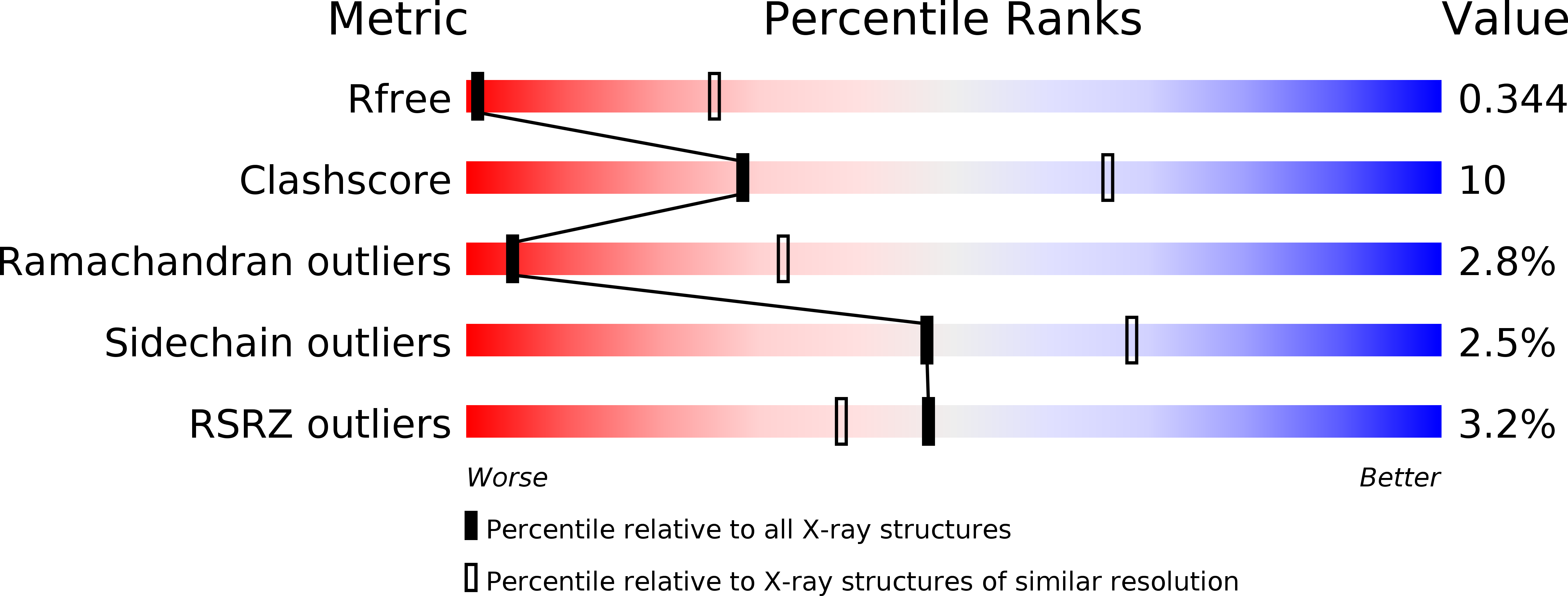
Resolution:
4.84 Å
R-Value Free:
0.35
R-Value Work:
0.33
R-Value Observed:
0.34
Space Group:
P 21 21 21