4AAY image
Entry Detail
PDB ID:
4AAY
Keywords:
Title:
Crystal Structure of the arsenite oxidase protein complex from Rhizobium species strain NT-26
Biological Source:
Host Organism:
PDB Version:
Deposition Date:
2011-12-05
Release Date:
2012-12-12
Method Details:
Experimental Method:
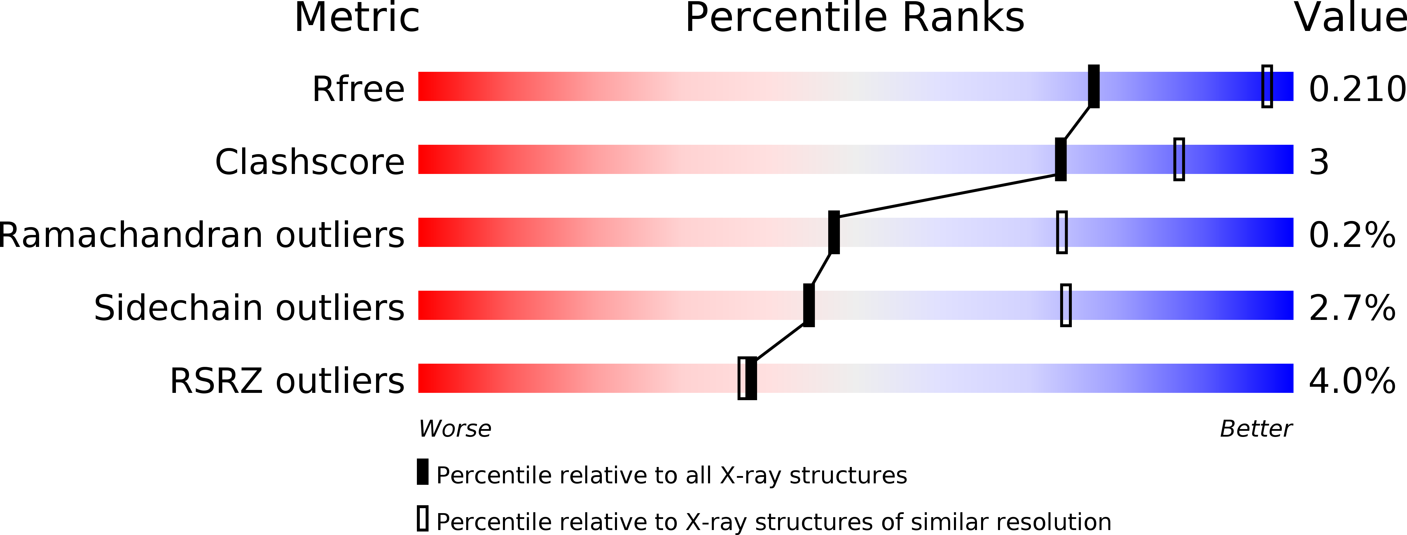
Resolution:
2.70 Å
R-Value Free:
0.21
R-Value Work:
0.19
R-Value Observed:
0.19
Space Group:
P 21 21 2
Macromolecular Entities
Polymer Type:polypeptide(L)
Description:AROA
Chain IDs:A, C, E, G
Chain Length:845
Number of Molecules:4
Biological Source:ARSENITE-OXIDISING BACTERIUM NT-26
Polymer Type:polypeptide(L)
Description:AROB
Chain IDs:B, D, F, H
Chain Length:175
Number of Molecules:4
Biological Source:ARSENITE-OXIDISING BACTERIUM NT-26
Primary Citation
The Respiratory Arsenite Oxidase: Structure and the Role of Residues Surrounding the Rieske Cluster.
Plos One 8 72535 ? (2013)
PMID: 24023621 DOI: 10.1371/JOURNAL.PONE.0072535

Abstact

The arsenite oxidase (Aio) from the facultative autotrophic Alphaproteobacterium Rhizobium sp. NT-26 is a bioenergetic enzyme involved in the oxidation of arsenite to arsenate. The enzyme from the distantly related heterotroph, Alcaligenes faecalis, which is thought to oxidise arsenite for detoxification, consists of a large α subunit (AioA) with bis-molybdopterin guanine dinucleotide at its active site and a 3Fe-4S cluster, and a small β subunit (AioB) which contains a Rieske 2Fe-2S cluster. The successful heterologous expression of the NT-26 Aio in Escherichia coli has resulted in the solution of its crystal structure. The NT-26 Aio, a heterotetramer, shares high overall similarity to the heterodimeric arsenite oxidase from A. faecalis but there are striking differences in the structure surrounding the Rieske 2Fe-2S cluster which we demonstrate explains the difference in the observed redox potentials (+225 mV vs. +130/160 mV, respectively). A combination of site-directed mutagenesis and electron paramagnetic resonance was used to explore the differences observed in the structure and redox properties of the Rieske cluster. In the NT-26 AioB the substitution of a serine (S126 in NT-26) for a threonine as in the A. faecalis AioB explains a -20 mV decrease in redox potential. The disulphide bridge in the A. faecalis AioB which is conserved in other betaproteobacterial AioB subunits and the Rieske subunit of the cytochrome bc 1 complex is absent in the NT-26 AioB subunit. The introduction of a disulphide bridge had no effect on Aio activity or protein stability but resulted in a decrease in the redox potential of the cluster. These results are in conflict with previous data on the betaproteobacterial AioB subunit and the Rieske of the bc 1 complex where removal of the disulphide bridge had no effect on the redox potential of the former but a decrease in cluster stability was observed in the latter.

Legend

Protein

Chemical

Disease

Primary Citation of related structures