4AAI image
Entry Detail
PDB ID:
4AAI
Keywords:
Title:
THERMOSTABLE PROTEIN FROM HYPERTHERMOPHILIC VIRUS SSV-RH
Biological Source:
Host Organism:
PDB Version:
Deposition Date:
2011-12-02
Release Date:
2012-01-11
Method Details:
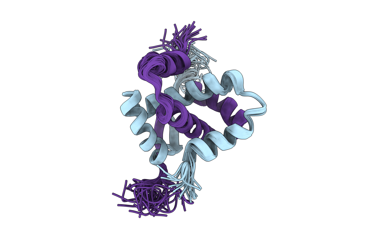
Experimental Method:
Conformers Calculated:
100
Conformers Submitted:
20
Selection Criteria:
LEAST RESTRAINT VIOLATION
Macromolecular Entities
Polymer Type:polypeptide(L)
Description:ORF E73
Chain IDs:A, B
Chain Length:73
Number of Molecules:2
Biological Source:SULFOLOBUS VIRUS RAGGED HILLS
Ligand Molecules
Primary Citation
Structural Studies of E73 from a Hyperthermophilic Archaeal Virus Identify the "Rh3" Domain, an Elaborated Ribbon-Helix- Helix Motif Involved in DNA Recognition.
Biochemistry 51 2899 ? (2012)
PMID: 22409376 DOI: 10.1021/BI201791S

Abstact

Hyperthermophilic archaeal viruses, including Sulfolobus spindle-shaped viruses (SSVs) such as SSV-1 and SSV-Ragged Hills, exhibit remarkable morphology and genetic diversity. However, they remain poorly understood, in part because their genomes exhibit limited or unrecognizable sequence similarity to genes with known function. Here we report structural and functional studies of E73, a 73-residue homodimeric protein encoded within the SSV-Ragged Hills genome. Despite lacking significant sequence similarity, the nuclear magnetic resonance (NMR) structure reveals clear similarity to ribbon-helix-helix (RHH) domains present in numerous proteins involved in transcriptional regulation. In vitro double-stranded DNA (dsDNA) binding experiments confirm the ability of E73 to bind dsDNA in a nonspecific manner with micromolar affinity, and characterization of the K11E variant confirms the location of the predicted DNA binding surface. E73 is distinct, however, from known RHH domains. The RHH motif is elaborated upon by the insertion of a third helix that is tightly integrated into the structural domain, giving rise to the "RH3" fold. Within the homodimer, this helix results in the formation of a conserved, symmetric cleft distal to the DNA binding surface, where it may mediate protein-protein interactions or contribute to the high thermal stability of E73. Analysis of backbone amide dynamics by NMR provides evidence of a rigid core, fast picosecond to nanosecond time scale NH bond vector motions for residues located within the antiparallel β-sheet region of the proposed DNA-binding surface, and slower microsecond to millisecond time scale motions for residues in the α1-α2 loop. The roles of E73 and its SSV homologues in the viral life cycle are discussed.

Legend

Protein

Chemical

Disease

Primary Citation of related structures