Deposition Date
2011-11-15
Release Date
2012-06-27
Last Version Date
2023-12-20
Entry Detail
PDB ID:
4A7W
Keywords:
Title:
Crystal structure of uridylate kinase from Helicobacter pylori
Biological Source:
Source Organism(s):
HELICOBACTER PYLORI (Taxon ID: 85962)
Expression System(s):
Method Details:
Experimental Method:
Resolution:
1.80 Å
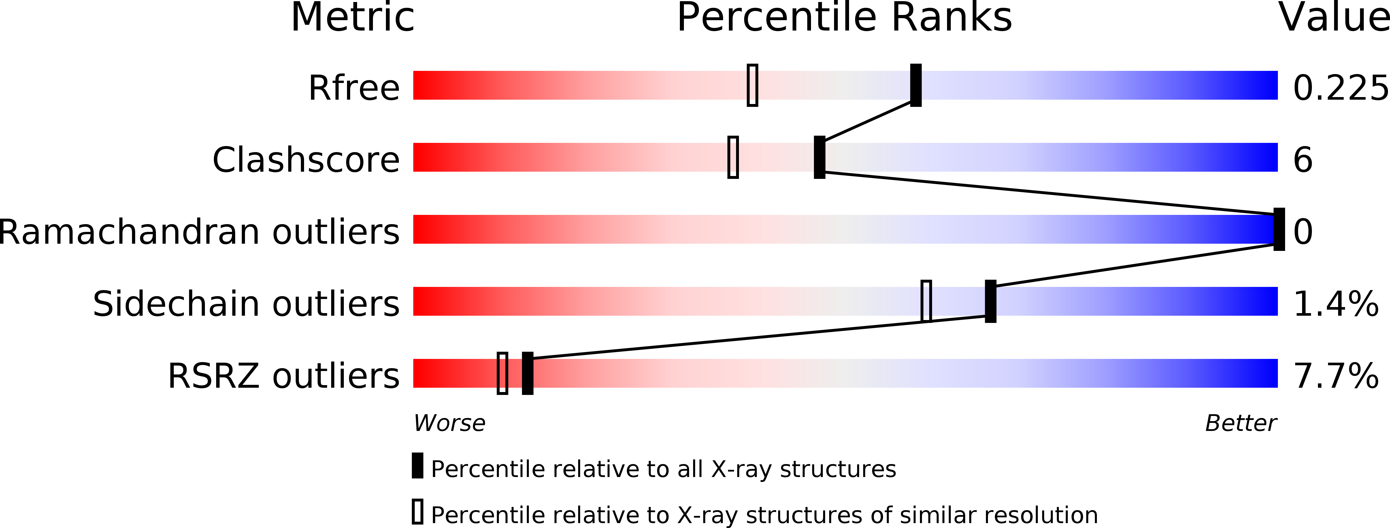
R-Value Free:
0.22
R-Value Work:
0.18
R-Value Observed:
0.19
Space Group:
H 3 2