Deposition Date
2011-11-08
Release Date
2012-01-25
Last Version Date
2023-12-20
Entry Detail
PDB ID:
4A6T
Keywords:
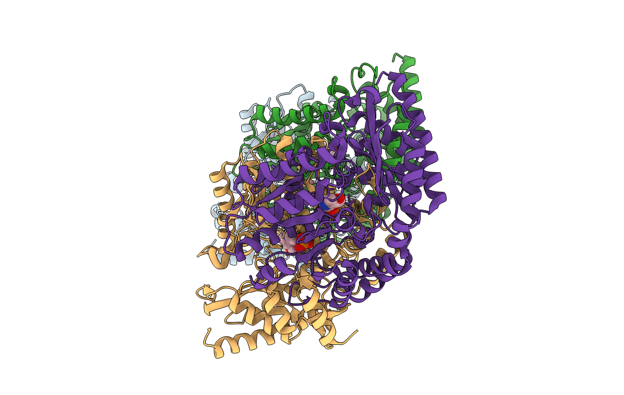
Title:
Crystal structure of the omega transaminase from Chromobacterium violaceum in complex with PLP
Biological Source:
Source Organism(s):
CHROMOBACTERIUM VIOLACEUM (Taxon ID: 536)
Expression System(s):
Method Details:
Experimental Method:
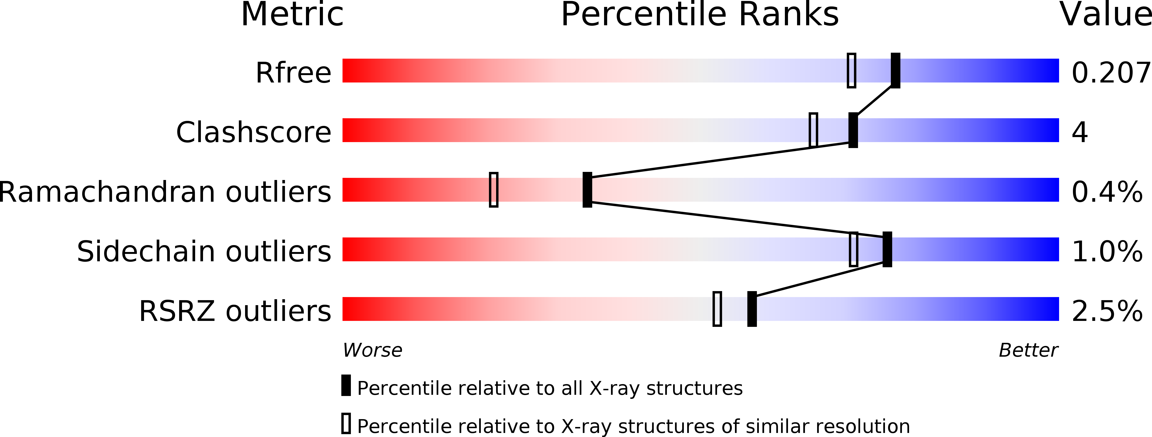
Resolution:
1.80 Å
R-Value Free:
0.20
R-Value Work:
0.16
R-Value Observed:
0.16
Space Group:
P 1