Deposition Date
2011-11-07
Release Date
2012-11-21
Last Version Date
2023-12-20
Entry Detail
PDB ID:
4A6N
Keywords:
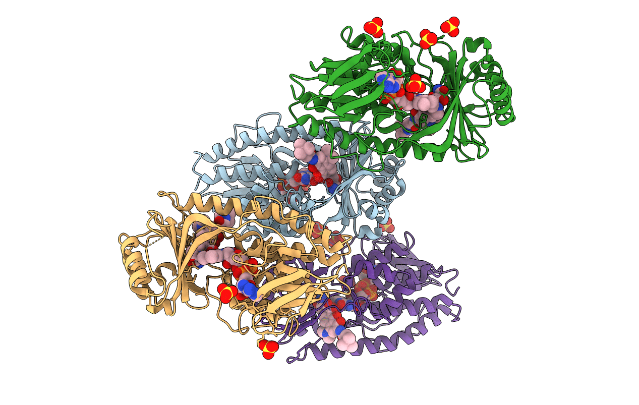
Title:
STRUCTURE OF THE TETRACYCLINE DEGRADING MONOOXYGENASE TETX IN COMPLEX WITH TIGECYCLINE
Biological Source:
Source Organism(s):
BACTEROIDES THETAIOTAOMICRON (Taxon ID: 818)
Expression System(s):
Method Details:
Experimental Method:
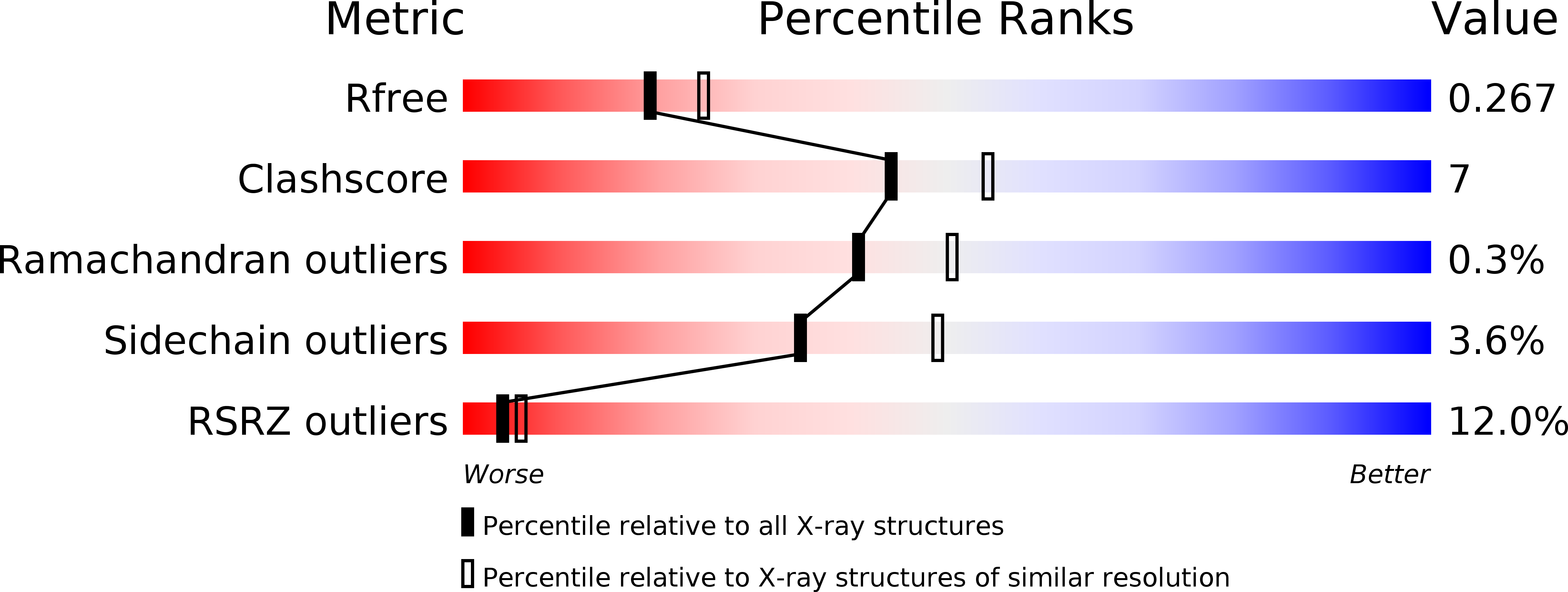
Resolution:
2.30 Å
R-Value Free:
0.26
R-Value Work:
0.21
R-Value Observed:
0.22
Space Group:
P 1