4A6F image
Entry Detail
PDB ID:
4A6F
Title:
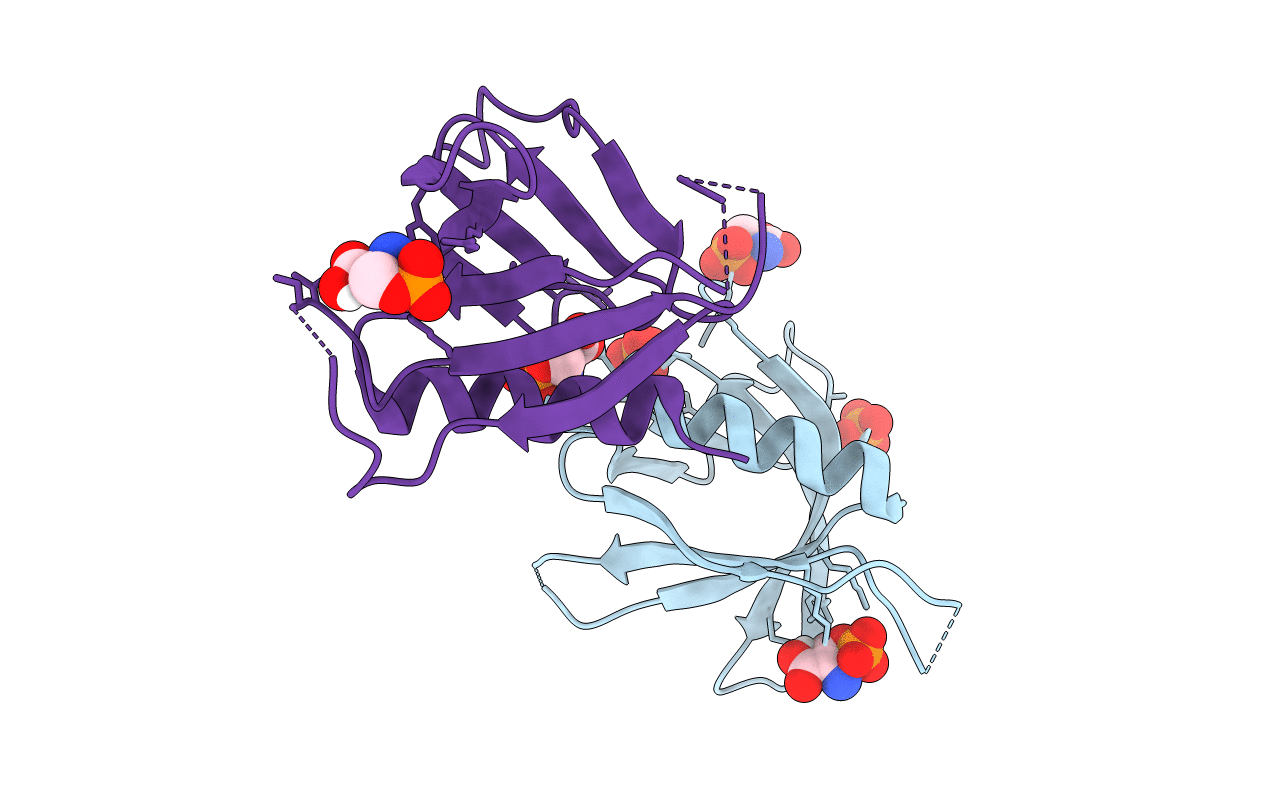
Crystal structure of Slm1-PH domain in complex with Phosphoserine
Biological Source:
Source Organism:
Host Organism:
PDB Version:
Deposition Date:
2011-11-02
Release Date:
2012-06-13
Method Details:
Experimental Method:
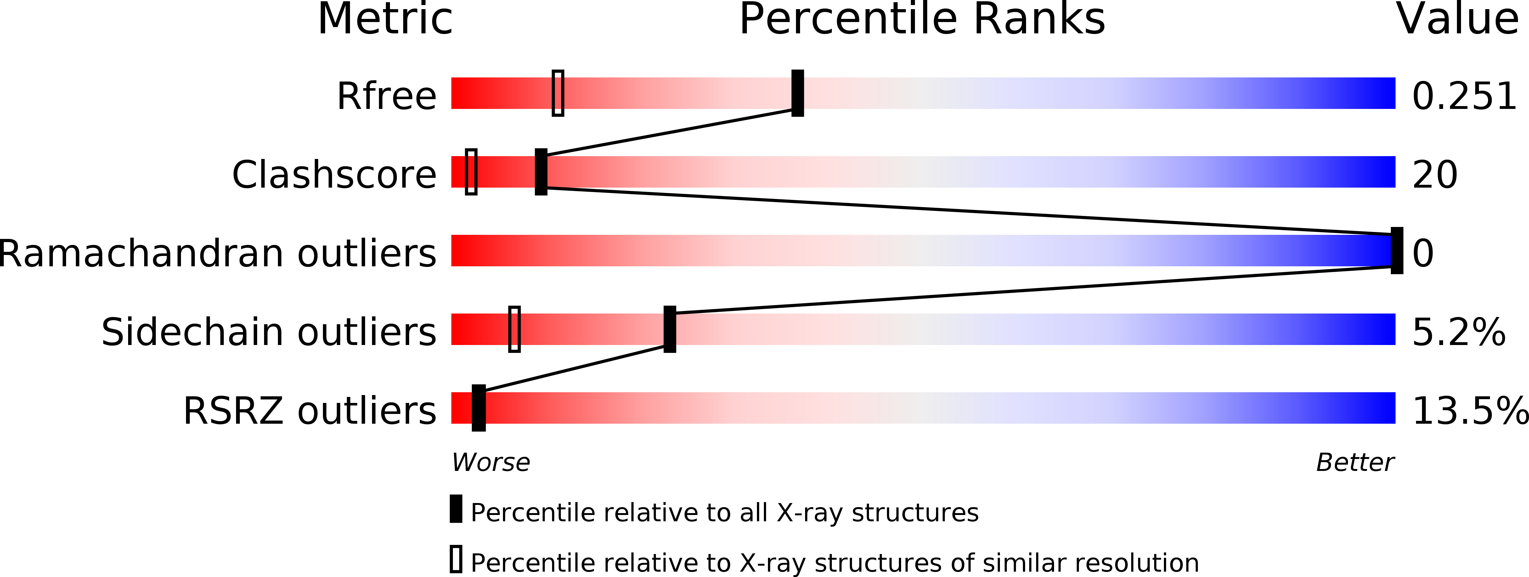
Resolution:
1.68 Å
R-Value Free:
0.24
R-Value Work:
0.21
R-Value Observed:
0.21
Space Group:
P 2 21 21
Macromolecular Entities
Polymer Type:polypeptide(L)
Description:PHOSPHATIDYLINOSITOL 4,5-BISPHOSPHATE-BINDING PROTEIN SLM1
Chain IDs:A
Chain Length:120
Number of Molecules:1
Biological Source:SACCHAROMYCES CEREVISIAE
Polymer Type:polypeptide(L)
Description:PHOSPHATIDYLINOSITOL 4,5-BISPHOSPHATE-BINDING PROTEIN SLM1
Chain IDs:B
Chain Length:120
Number of Molecules:1
Biological Source:SACCHAROMYCES CEREVISIAE
Primary Citation
Structural Analyses of Slm1-Ph Domain Demonstrate Ligand Binding in the Non-Canonical Site
Plos One 7 36526 ? (2012)
PMID: 22574179 DOI: 10.1371/JOURNAL.PONE.0036526

Abstact

BACKGROUND Pleckstrin homology (PH) domains are common membrane-targeting modules and their best characterized ligands are a set of important signaling lipids that include phosphatidylinositol phosphates (PtdInsPs). PH domains recognize PtdInsPs through two distinct mechanisms that use different binding pockets on opposite sides of the β-strands 1 and 2: i) a canonical binding site delimited by the β1-β2 and β3-β4loops and ii) a non-canonical binding site bordered by the β1-β2 and β5-β6loops. The PH domain-containing protein Slm1 from budding yeast Saccharomyces cerevisiae is required for actin cytoskeleton polarization and cell growth. We recently reported that this PH domain binds PtdInsPs and phosphorylated sphingolipids in a cooperative manner. PRINCIPAL FINDINGS To study the structural basis for the Slm1-PH domain (Slm1-PH) specificity, we co-crystallized this domain with different soluble compounds that have structures analogous to anionic lipid head groups of reported Slm1 ligands: inositol 4-phosphate, which mimics phosphatidylinositol-4-phosphate (PtdIns(4)P), and phosphoserine as a surrogate for dihydrosphingosine 1-phosphate (DHS1-P). We found electron densities for the ligands within the so-called non-canonical binding site. An additional positively charged surface that contacts a phosphate group was identified next to the canonical binding site. CONCLUSIONS Our results suggest that Slm1-PH utilizes a non-canonical binding site to bind PtdInsPs, similar to that described for the PH domains of β-spectrin, Tiam1 and ArhGAP9. Additionally, Slm1-PH may have retained an active canonical site. We propose that the presence of both a canonical and a non-canonical binding pocket in Slm1-PH may account for the cooperative binding to PtdInsPs and DHS-1P.

Legend

Protein

Chemical

Disease

Primary Citation of related structures