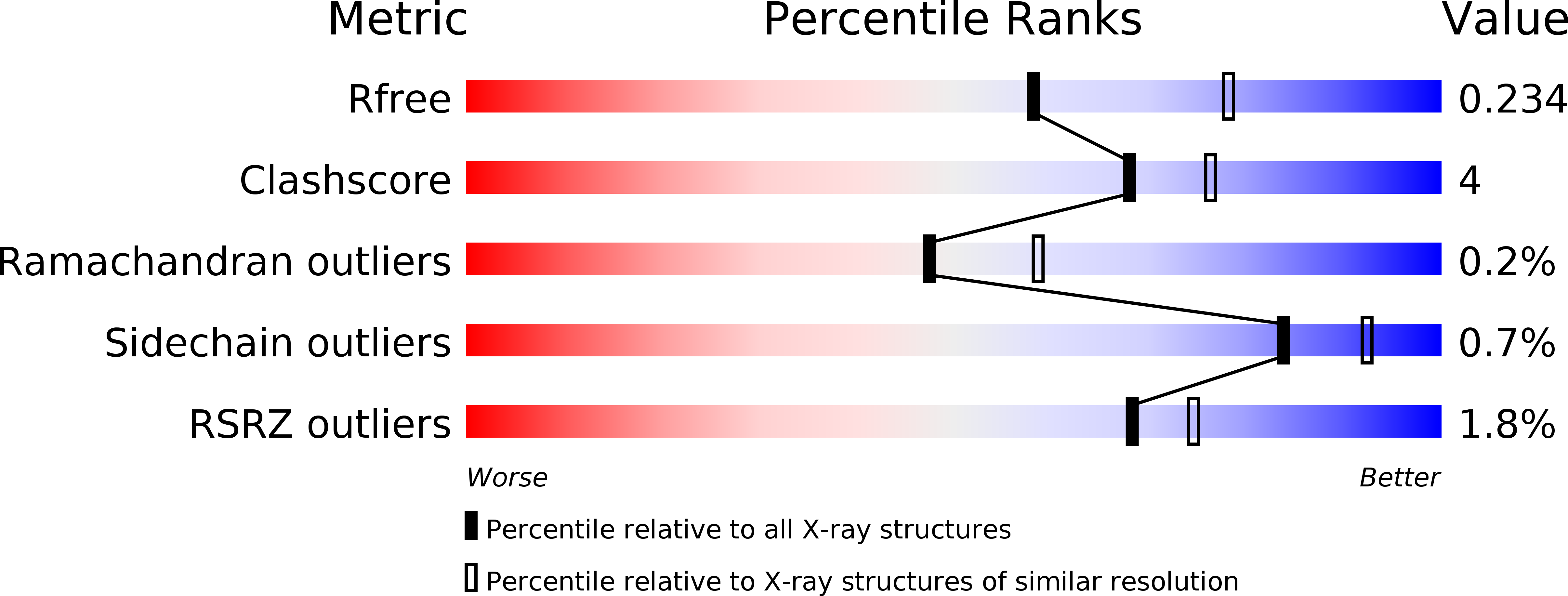
4A5Z image
Deposition Date 2011-10-30
Release Date 2012-11-14
Last Version Date 2024-10-23
Entry Detail
PDB ID:
4A5Z
Title:
Structures of MITD1
Biological Source:
Source Organism:
HOMO SAPIENS (Taxon ID: 9606)
Host Organism:
Method Details:
Experimental Method:
Resolution:
2.30 Å
R-Value Free:
0.22
R-Value Work:
0.19
R-Value Observed:
0.19
Space Group:
P 1 21 1
Macromolecular Entities
Structures with similar UniProt ID
Protein Blast
Polymer Type:polypeptide(L)
Molecule:MIT DOMAIN-CONTAINING PROTEIN 1
Gene (Uniprot):MITD1
Mutagens:YES
Chain IDs:A, B, C, D
Chain Length:163
Number of Molecules:4
Biological Source:HOMO SAPIENS
Modified Residue
Compound ID Chain ID Parent Comp ID Details 2D Image
MSE A MET SELENOMETHIONINE
Primary Citation
Escrt-III Binding Protein Mitd1 is Involved in Cytokinesis and Has an Unanticipated Pld Fold that Binds Membranes.
Proc.Natl.Acad.Sci.USA 109 17424 ? (2012)
PMID: 23045692 DOI: 10.1073/PNAS.1206839109

Abstact

The endosomal sorting complexes required for transport (ESCRT) proteins have a critical function in abscission, the final separation of the daughter cells during cytokinesis. Here, we describe the structure and function of a previously uncharacterized ESCRT-III interacting protein, MIT-domain containing protein 1 (MITD1). Crystal structures of MITD1 reveal a dimer, with a microtubule-interacting and trafficking (MIT) domain at the N terminus and a unique, unanticipated phospholipase D-like (PLD) domain at the C terminus that binds membranes. We show that the MIT domain binds to a subset of ESCRT-III subunits and that this interaction mediates MITD1 recruitment to the midbody during cytokinesis. Depletion of MITD1 causes a distinct cytokinetic phenotype consistent with destabilization of the midbody and abscission failure. These results suggest a model whereby MITD1 coordinates the activity of ESCRT-III during abscission with earlier events in the final stages of cell division.

Legend

Protein

Chemical

Disease

Primary Citation of related structures