4A3Q image
Deposition Date 2011-10-04
Release Date 2011-12-28
Last Version Date 2023-12-20
Entry Detail
PDB ID:
4A3Q
Keywords:
Title:
The 2.15 Angstrom resolution crystal structure of Staphylococcus aureus alanine racemase
Biological Source:
Source Organism:
Host Organism:
Method Details:
Experimental Method:
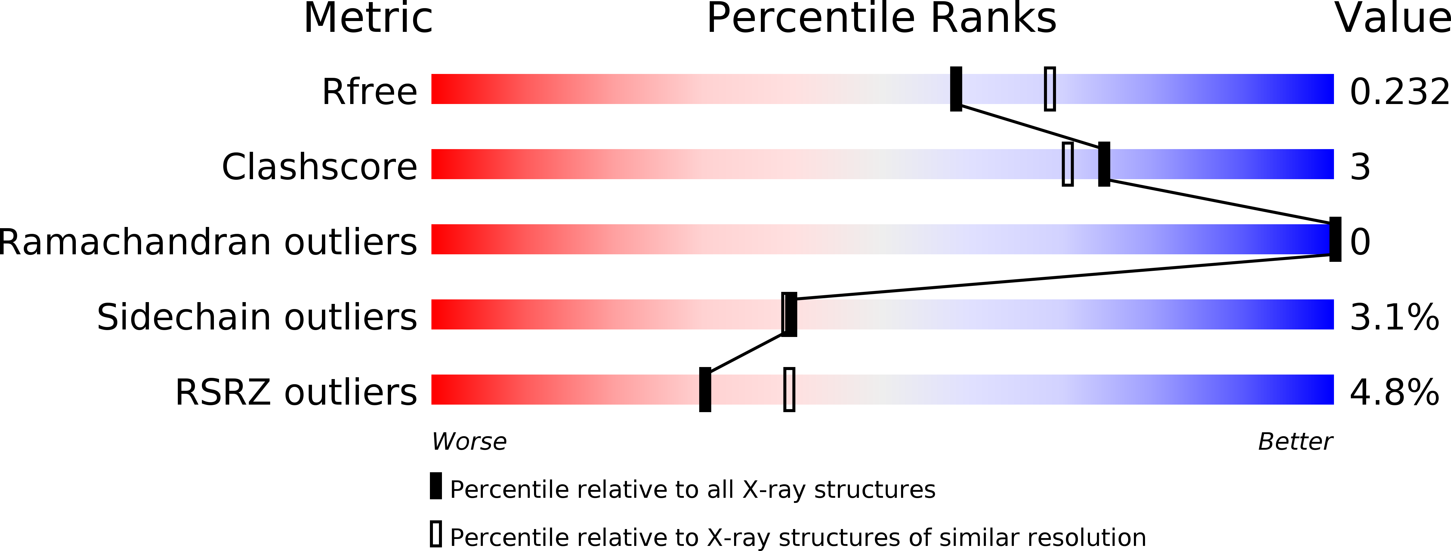
Resolution:
2.15 Å
R-Value Free:
0.23
R-Value Work:
0.18
R-Value Observed:
0.19
Space Group:
P 21 21 21
Macromolecular Entities
Structures with similar UniProt ID
Protein Blast
Polymer Type:polypeptide(L)
Molecule:ALANINE RACEMASE 1
Gene (Uniprot):alr1
Chain IDs:A, B
Chain Length:382
Number of Molecules:2
Biological Source:STAPHYLOCOCCUS AUREUS
Primary Citation
Structural Features and Kinetic Characterization of Alanine Racemase from Staphylococcus Aureus (Mu50)
Acta Crystallogr.,Sect.D 68 82 ? (2012)
PMID: 22194336 DOI: 10.1107/S0907444911050682

Abstact

Staphylococcus aureus is an opportunistic Gram-positive bacterium which causes a wide variety of diseases ranging from minor skin infections to potentially fatal conditions such as pneumonia, meningitis and septicaemia. The pathogen is a leading cause of nosocomial acquired infections, a problem that is exacerbated by the existence of methicillin- and glycopeptide antibiotic-resistant strains which can be challenging to treat. Alanine racemase (Alr) is a pyridoxal-5'-phosphate-dependent enzyme which catalyzes reversible racemization between enantiomers of alanine. As D-alanine is an essential component of the bacterial cell-wall peptidoglycan, inhibition of Alr is lethal to prokaryotes. Additionally, while ubiquitous amongst bacteria, this enzyme is absent in humans and most eukaryotes, making it an excellent antibiotic drug target. The crystal structure of S. aureus alanine racemase (Alr(Sas)), the sequence of which corresponds to that from the highly antibiotic-resistant Mu50 strain, has been solved to 2.15 Å resolution. Comparison of the Alr(Sas) structure with those of various alanine racemases demonstrates a conserved overall fold, with the enzyme sharing most similarity to those from other Gram-positive bacteria. Structural examination indicates that the active-site binding pocket, dimer interface and active-site entryway of the enzyme are potential targets for structure-aided inhibitor design. Kinetic constants were calculated in this study and are reported here. The potential for a disulfide bond in this structure is noted. This structural and biochemical information provides a template for future structure-based drug-development efforts targeting Alr(Sas).

Legend

Protein

Chemical

Disease

Primary Citation of related structures
Feedback Form
Name
Email
Institute
Feedback