Deposition Date
2011-09-28
Release Date
2012-06-27
Last Version Date
2024-10-23
Entry Detail
PDB ID:
4A2O
Keywords:
Title:
STRUCTURE OF THE HUMAN EOSINOPHIL CATIONIC PROTEIN IN COMPLEX WITH SULFATE ANIONS
Biological Source:
Source Organism(s):
HOMO SAPIENS (Taxon ID: 9606)
Expression System(s):
Method Details:
Experimental Method:
Resolution:
1.69 Å
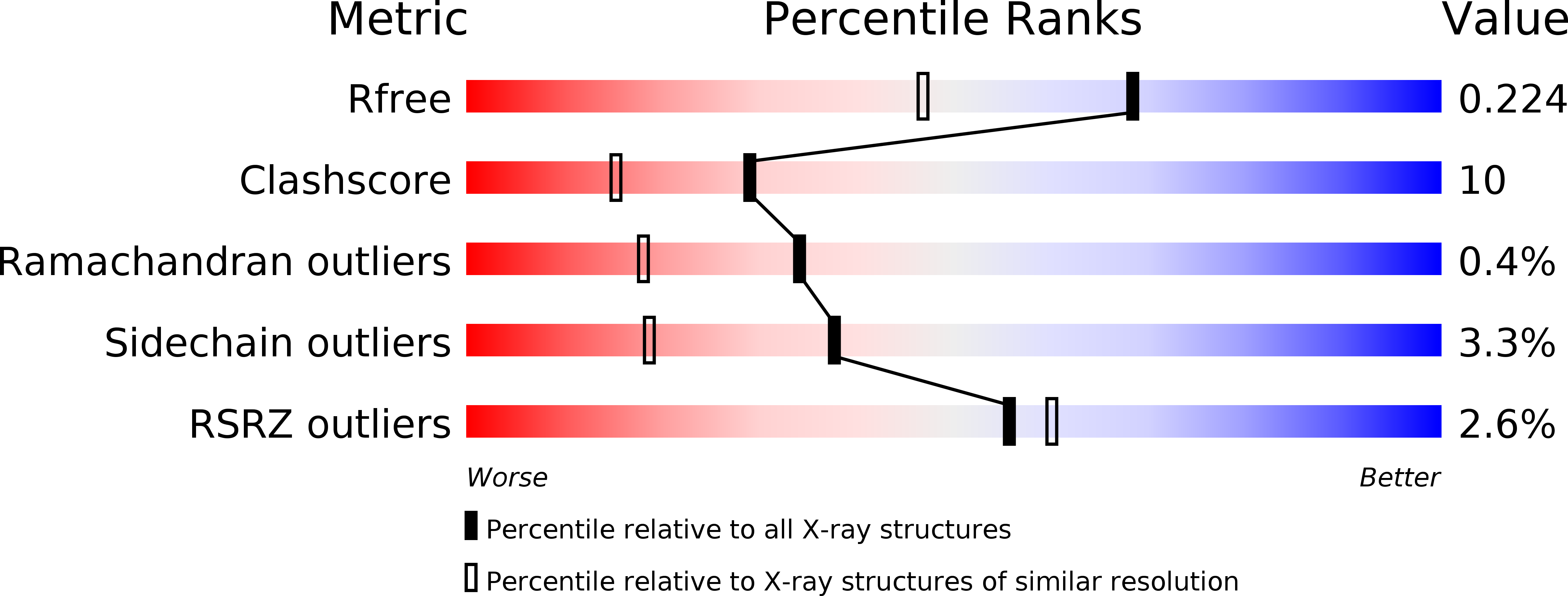
R-Value Free:
0.22
R-Value Work:
0.16
R-Value Observed:
0.16
Space Group:
C 1 2 1