4A2C image
Deposition Date 2011-09-26
Release Date 2012-08-08
Last Version Date 2023-12-20
Entry Detail
PDB ID:
4A2C
Keywords:
Title:
Crystal structure of galactitol-1-phosphate dehydrogenase from Escherichia coli
Biological Source:
Source Organism:
Host Organism:
Method Details:
Experimental Method:
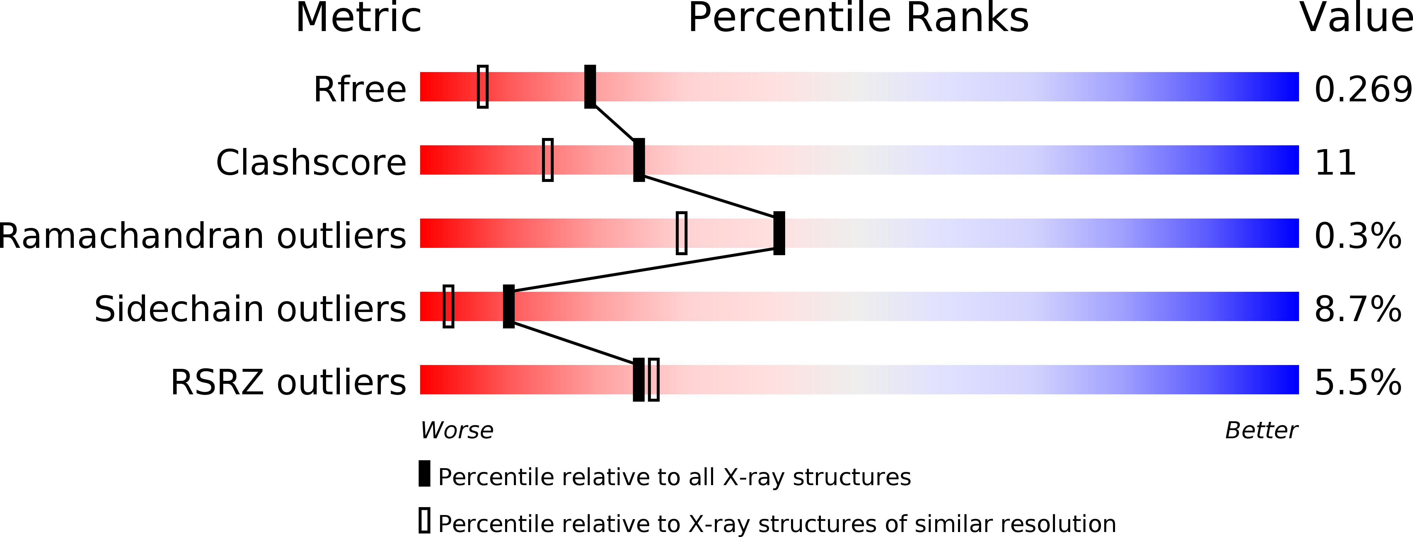
Resolution:
1.87 Å
R-Value Free:
0.27
R-Value Work:
0.21
R-Value Observed:
0.21
Space Group:
P 1 21 1
Macromolecular Entities
Structures with similar UniProt ID
Protein Blast
Polymer Type:polypeptide(L)
Molecule:GALACTITOL-1-PHOSPHATE 5-DEHYDROGENASE
Gene (Uniprot):gatD
Chain IDs:A, B
Chain Length:346
Number of Molecules:2
Biological Source:ESCHERICHIA COLI
Primary Citation
The Crystal Structure of Galactitol-1-Phosphate 5-Dehydrogenase from Escherichia Coli K12 Provides Insights Into its Anomalous Behavior on Imac Processes
FEBS Lett. 586 3127 ? (2012)
PMID: 22979983 DOI: 10.1016/J.FEBSLET.2012.07.073

Abstact

Endogenous galactitol-1-phosphate 5-dehydrogenase (GPDH) (EC 1.1.1.251) from Escherichia coli spontaneously interacts with Ni(2+)-NTA matrices becoming a potential contaminant for recombinant, target His-tagged proteins. Purified recombinant, untagged GPDH (rGPDH) converted galactitol into tagatose, and d-tagatose-6-phosphate into galactitol-1-phosphate, in a Zn(2+)- and NAD(H)-dependent manner and readily crystallized what has permitted to solve its crystal structure. In contrast, N-terminally His-tagged GPDH was marginally stable and readily aggregated. The structure of rGPDH revealed metal-binding sites characteristic from the medium-chain dehydrogenase/reductase protein superfamily which may explain its ability to interact with immobilized metals. The structure also provides clues on the harmful effects of the N-terminal His-tag.

Legend

Protein

Chemical

Disease

Primary Citation of related structures
Feedback Form
Name
Email
Institute
Feedback