4A1F image
Deposition Date 2011-09-14
Release Date 2012-03-21
Last Version Date 2024-05-08
Entry Detail
PDB ID:
4A1F
Keywords:
Title:
Crystal structure of C-terminal domain of Helicobacter pylori DnaB Helicase
Biological Source:
Source Organism:
Host Organism:
Method Details:
Experimental Method:
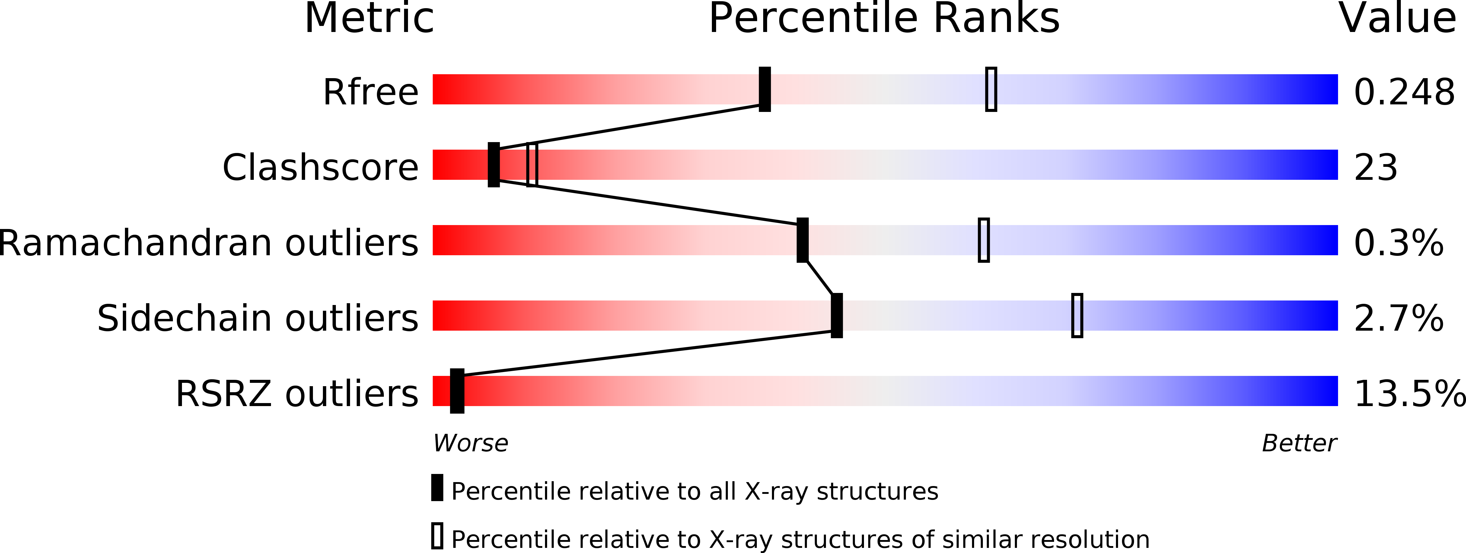
Resolution:
2.50 Å
R-Value Free:
0.25
R-Value Work:
0.21
R-Value Observed:
0.21
Space Group:
P 21 21 2
Macromolecular Entities
Polymer Type:polypeptide(L)
Molecule:REPLICATIVE DNA HELICASE
Gene (Uniprot):dnaB
Chain IDs:A, B
Chain Length:338
Number of Molecules:2
Biological Source:HELICOBACTER PYLORI
Ligand Molecules
Primary Citation
Architecture of a Dodecameric Bacterial Replicative Helicase.
Structure 20 554 ? (2012)
PMID: 22405014 DOI: 10.1016/J.STR.2012.01.020

Abstact

Hexameric DnaB helicases are often loaded at DNA replication forks by interacting with the initiator protein DnaA and/or a helicase loader (DnaC in Escherichia coli). These loaders are not universally required, and DnaB from Helicobacter pylori was found to bypass DnaC when expressed in E. coli cells. The crystal structure of Helicobacter pylori DnaB C-terminal domain (HpDnaB-CTD) reveals a large two-helix insertion (named HPI) in the ATPase domain that protrudes away from the RecA fold. Biophysical characterization and electron microscopy (EM) analysis of the full-length protein show that HpDnaB forms head-to-head double hexamers remarkably similar to helicases found in some eukaryotes, archaea, and viruses. The docking of the HpDnaB-CTD structure into EM reconstruction of HpDnaB provides a model that shows how hexamerization of the CTD is facilitated by HPI-HPI interactions. The HpDnaB double-hexamer architecture supports an alternative strategy to load bacterial helicases onto forks in the absence of helicase loaders.

Legend

Protein

Chemical

Disease

Primary Citation of related structures