
Deposition Date
1998-06-10
Release Date
1998-07-06
Last Version Date
2024-02-28
Entry Detail
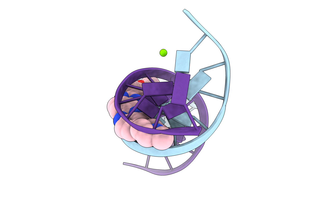
PDB ID:
403D
Keywords:
Title:
5'-D(*CP*GP*CP*(HYD)AP*AP*AP*TP*TP*TP*GP*CP*G)-3', 2'-(4-ETHOXYPHENYL)-5-(4-METHYL-1-PIPERAZINYL)-2,5'-BI-BENZIMIDAZOLE
Method Details:
Experimental Method:
Resolution:
1.40 Å
R-Value Free:
0.31
R-Value Work:
0.24
R-Value Observed:
0.24
Space Group:
P 21 21 21
