Deposition Date
2011-07-28
Release Date
2011-11-09
Last Version Date
2024-05-08
Entry Detail
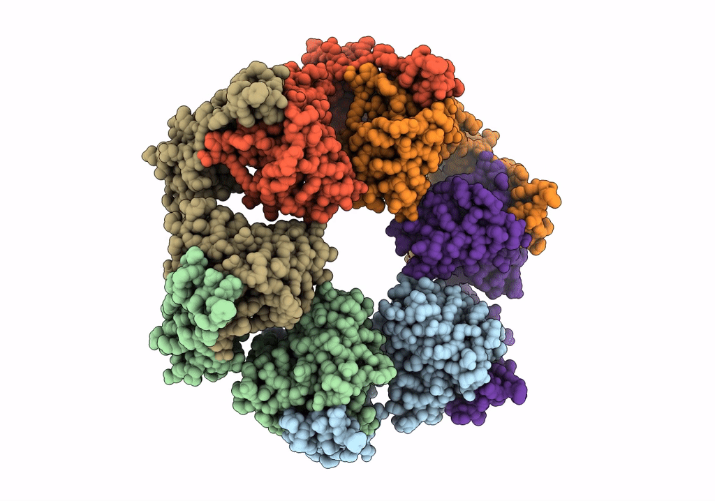
PDB ID:
3ZW6
Keywords:
Title:
MODEL OF HEXAMERIC AAA DOMAIN ARRANGEMENT OF GREEN-TYPE RUBISCO ACTIVASE FROM TOBACCO.
Biological Source:
Source Organism:
NICOTIANA TABACUM (Taxon ID: 4097)
Host Organism:
Method Details:
Experimental Method:
Resolution:
20.00 Å
Aggregation State:
PARTICLE
Reconstruction Method:
SINGLE PARTICLE