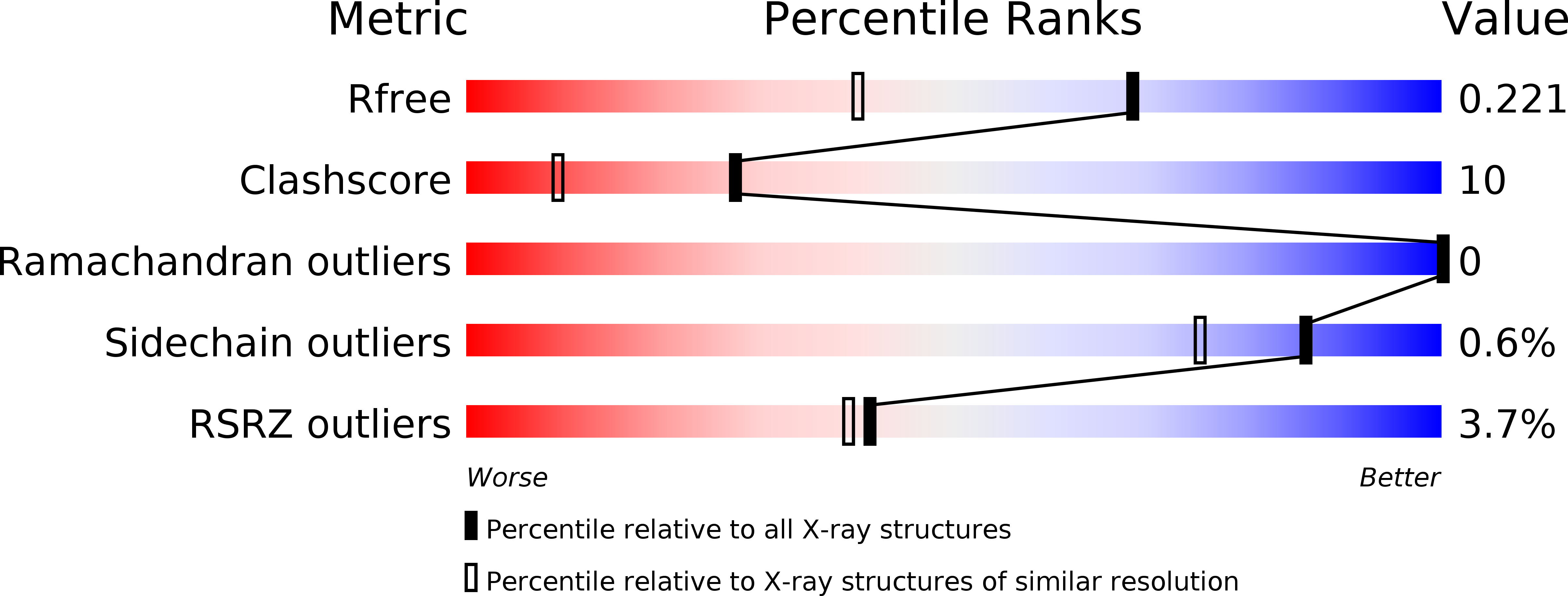
3sqf image
Deposition Date 2011-07-05
Release Date 2011-09-21
Last Version Date 2023-09-13
Entry Detail
PDB ID:
3SQF
Keywords:
Title:
Crystal structure of monomeric M-PMV retroviral protease
Biological Source:
Source Organism:
Host Organism:
Method Details:
Experimental Method:
Resolution:
1.63 Å
R-Value Free:
0.21
R-Value Work:
0.16
R-Value Observed:
0.17
Space Group:
P 1 21 1
Macromolecular Entities
Polymer Type:polypeptide(L)
Molecule:Protease
Gene (Uniprot):gag-pro
Mutations:C7A, C106A, D26N
Chain IDs:A, B
Chain Length:114
Number of Molecules:2
Biological Source:Mason-Pfizer monkey virus
Primary Citation

Abstact

Following the failure of a wide range of attempts to solve the crystal structure of M-PMV retroviral protease by molecular replacement, we challenged players of the protein folding game Foldit to produce accurate models of the protein. Remarkably, Foldit players were able to generate models of sufficient quality for successful molecular replacement and subsequent structure determination. The refined structure provides new insights for the design of antiretroviral drugs.

Legend

Protein

Chemical

Disease

Primary Citation of related structures