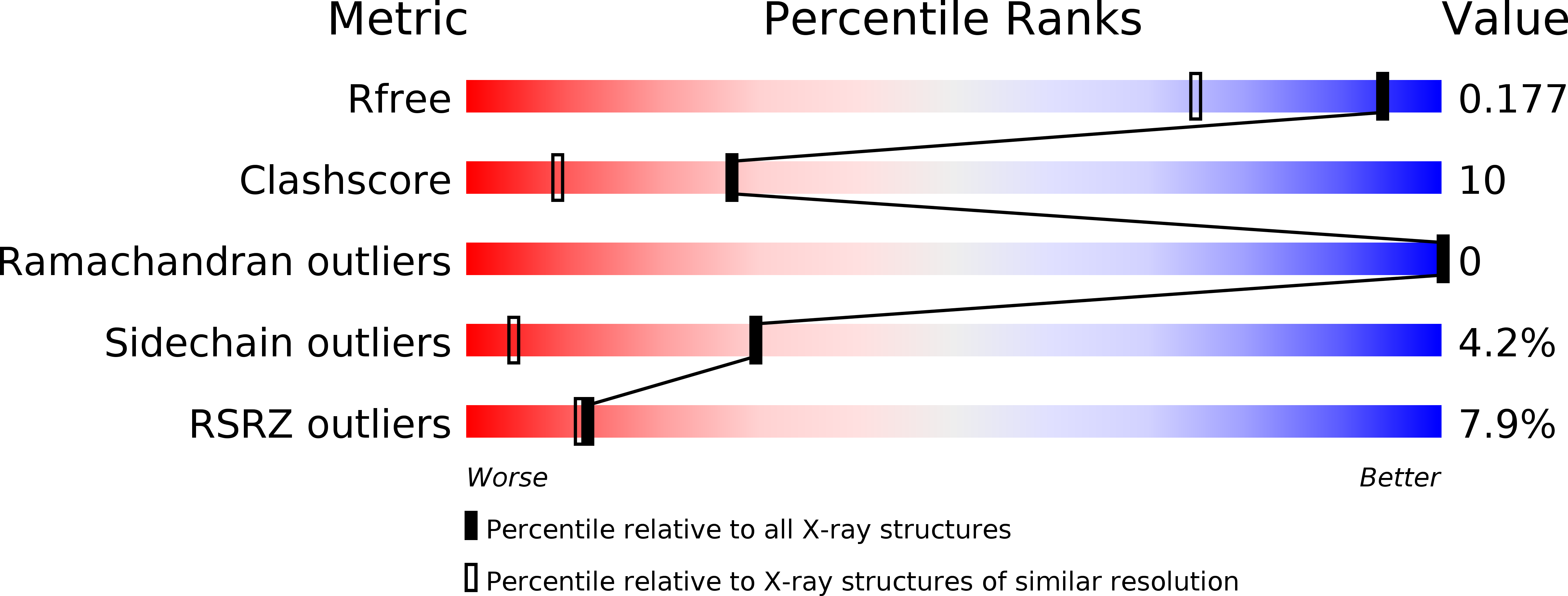
3slz image
Deposition Date 2011-06-27
Release Date 2011-10-12
Last Version Date 2024-02-28
Entry Detail
PDB ID:
3SLZ
Title:
The crystal structure of XMRV protease complexed with TL-3
Biological Source:
Source Organism:
Host Organism:
Method Details:
Experimental Method:
Resolution:
1.40 Å
R-Value Free:
0.20
R-Value Work:
0.17
R-Value Observed:
0.17
Space Group:
P 21 21 21
Macromolecular Entities
Structures with similar UniProt ID
Protein Blast
Polymer Type:polypeptide(L)
Molecule:gag-pro-pol polyprotein
Chain IDs:A, B
Chain Length:132
Number of Molecules:2
Biological Source:DG-75 Murine leukemia virus
Peptide-like Molecules
PRD_000434
Primary Citation
Structural and biochemical characterization of the inhibitor complexes of xenotropic murine leukemia virus-related virus protease.
Febs J. 278 4413 4424 (2011)
PMID: 21951660 DOI: 10.1111/j.1742-4658.2011.08364.x

Abstact

Interactions between the protease (PR) encoded by the xenotropic murine leukemia virus-related virus and a number of potential inhibitors have been investigated by biochemical and structural techniques. It was observed that several inhibitors used clinically against HIV PR exhibit nanomolar or even subnanomolar values of K(i) , depending on the exact experimental conditions. Both TL-3, a universal inhibitor of retroviral PRs, and some inhibitors originally shown to inhibit plasmepsins were also quite potent, whereas inhibition by pepstatin A was considerably weaker. Crystal structures of the complexes of xenotropic murine leukemia virus-related virus PR with TL-3, amprenavir and pepstatin A were solved at high resolution and compared with the structures of complexes of these inhibitors with other retropepsins. Whereas TL-3 and amprenavir bound in a predictable manner, spanning the substrate-binding site of the enzyme, two molecules of pepstatin A bound simultaneously in an unprecedented manner, leaving the catalytic water molecule in place.

Legend

Protein

Chemical

Disease

Primary Citation of related structures