Deposition Date
2010-02-23
Release Date
2010-03-23
Last Version Date
2024-11-20
Entry Detail
PDB ID:
3LW1
Keywords:
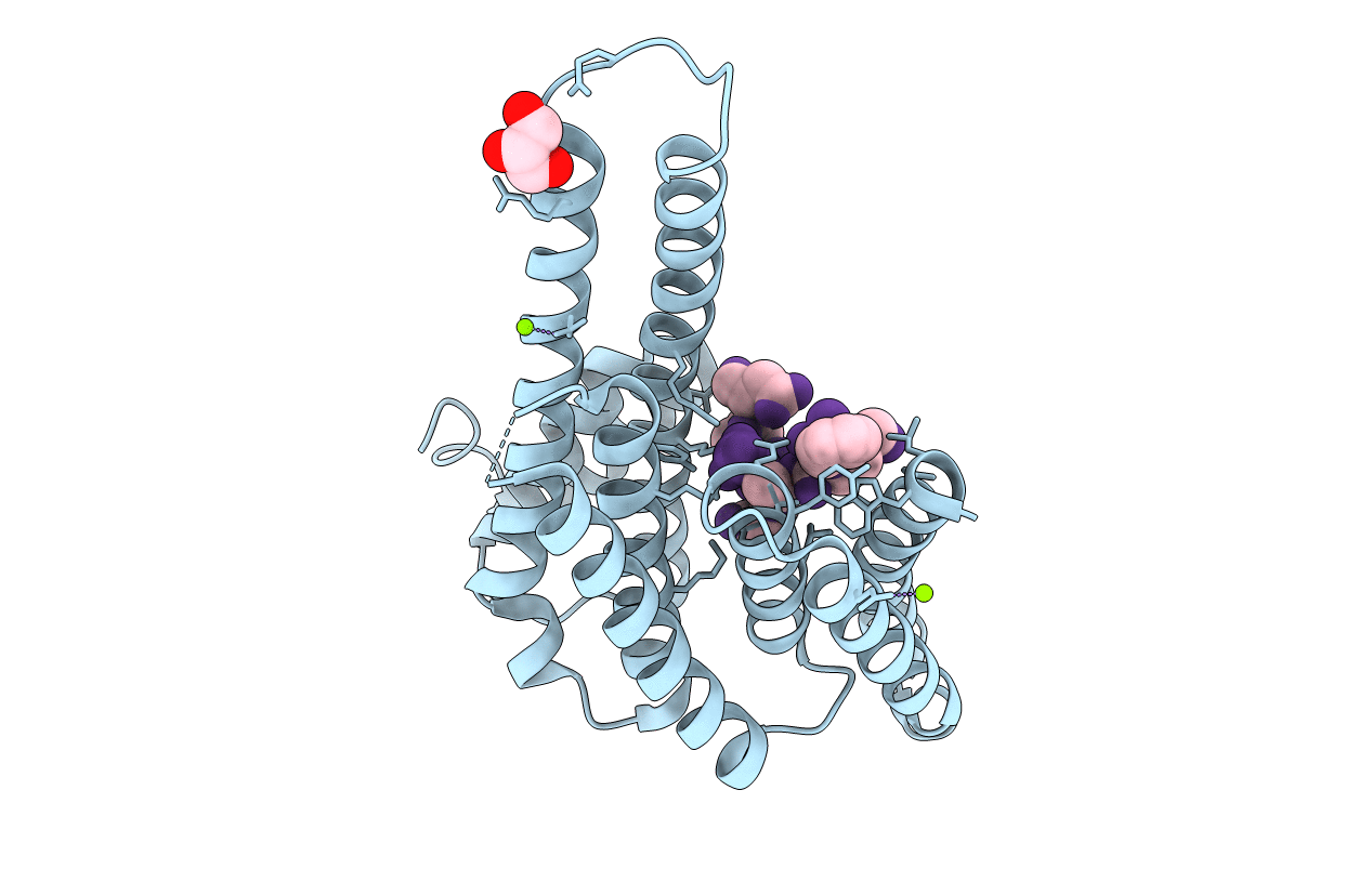
Title:
Binary complex of 14-3-3 sigma and p53 pT387-peptide
Biological Source:
Source Organism(s):
Homo sapiens (Taxon ID: 9606)
Expression System(s):
Method Details:
Experimental Method:
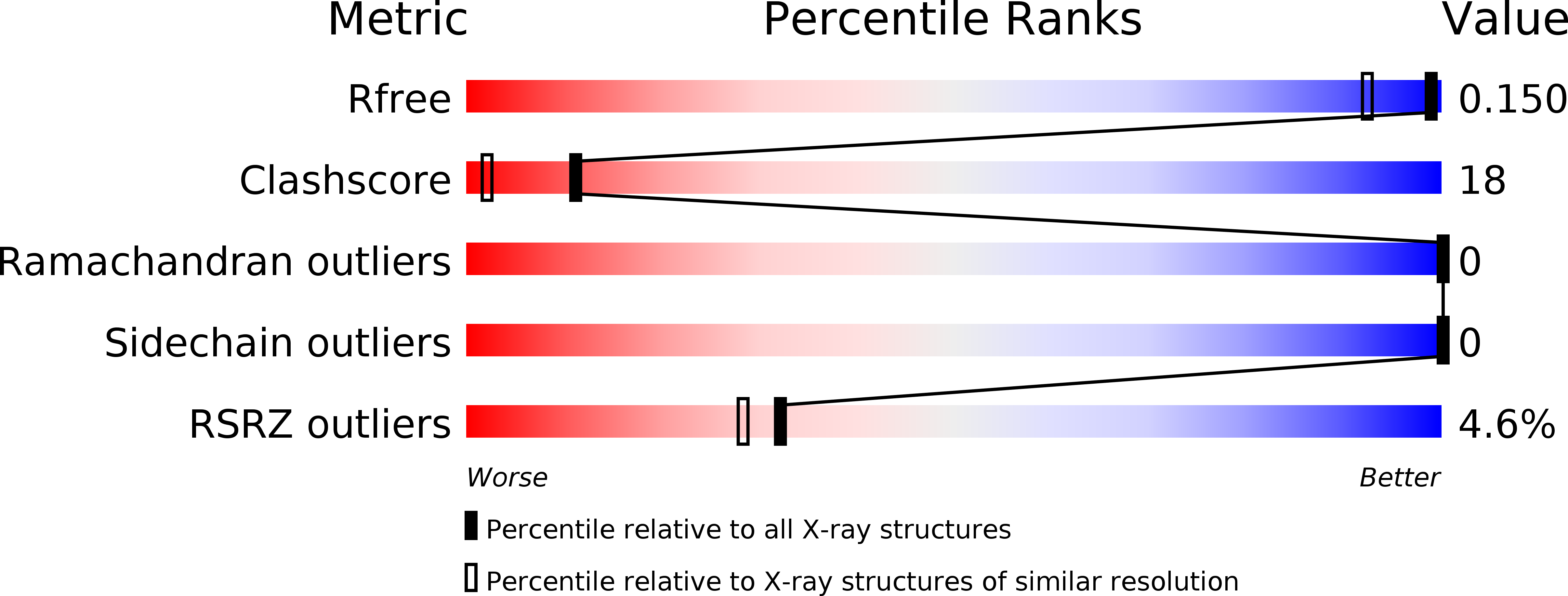
Resolution:
1.28 Å
R-Value Free:
0.15
R-Value Work:
0.12
R-Value Observed:
0.12
Space Group:
C 2 2 21