3lnt image
Deposition Date 2010-02-03
Release Date 2010-02-09
Last Version Date 2024-02-21
Entry Detail
PDB ID:
3LNT
Keywords:
Title:
Crystal structure of phosphoglyceromutase from Burkholderia Pseudomallei 1710B with bound malonic acid
Biological Source:
Source Organism:
Host Organism:
Method Details:
Experimental Method:
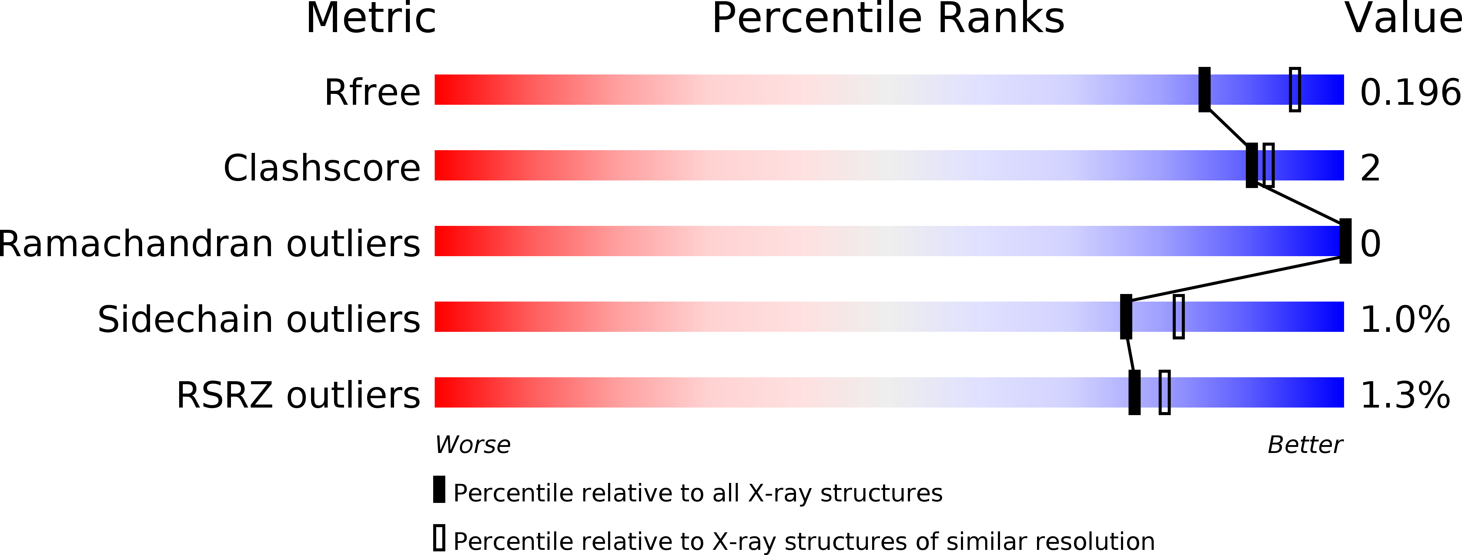
Resolution:
2.10 Å
R-Value Free:
0.19
R-Value Work:
0.17
R-Value Observed:
0.17
Space Group:
P 65
Macromolecular Entities
Polymer Type:polypeptide(L)
Molecule:2,3-bisphosphoglycerate-dependent phosphoglycerate mutase
Gene (Uniprot):gpmA
Chain IDs:A, B
Chain Length:250
Number of Molecules:2
Biological Source:Burkholderia pseudomallei
Primary Citation
An ensemble of structures of Burkholderia pseudomallei 2,3-bisphosphoglycerate-dependent phosphoglycerate mutase.
Acta Crystallogr.,Sect.F 67 1044 1050 (2011)
PMID: 21904048 DOI: 10.1107/S1744309111030405

Abstact

Burkholderia pseudomallei is a soil-dwelling bacterium endemic to Southeast Asia and Northern Australia. Burkholderia is responsible for melioidosis, a serious infection of the skin. The enzyme 2,3-bisphosphoglycerate-dependent phosphoglycerate mutase (PGAM) catalyzes the interconversion of 3-phosphoglycerate and 2-phosphoglycerate, a key step in the glycolytic pathway. As such it is an extensively studied enzyme and X-ray crystal structures of PGAM enzymes from multiple species have been elucidated. Vanadate is a phosphate mimic that is a powerful tool for studying enzymatic mechanisms in phosphoryl-transfer enzymes such as phosphoglycerate mutase. However, to date no X-ray crystal structures of phosphoglycerate mutase have been solved with vanadate acting as a substrate mimic. Here, two vanadate complexes together with an ensemble of substrate and fragment-bound structures that provide a comprehensive picture of the function of the Burkholderia enzyme are reported.

Legend

Protein

Chemical

Disease

Primary Citation of related structures