Deposition Date
2009-10-20
Release Date
2009-12-15
Last Version Date
2024-11-27
Entry Detail
PDB ID:
3KBH
Keywords:
Title:
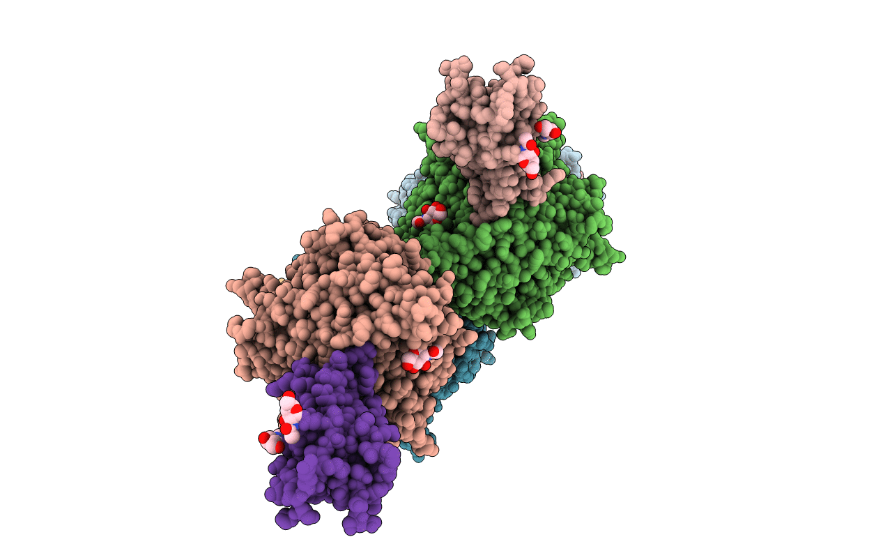
Crystal structure of NL63 respiratory coronavirus receptor-binding domain complexed with its human receptor
Biological Source:
Source Organism:
Homo sapiens (Taxon ID: 9606)
Human coronavirus NL63 (Taxon ID: 277944)
Human coronavirus NL63 (Taxon ID: 277944)
Host Organism:
Method Details:
Experimental Method:
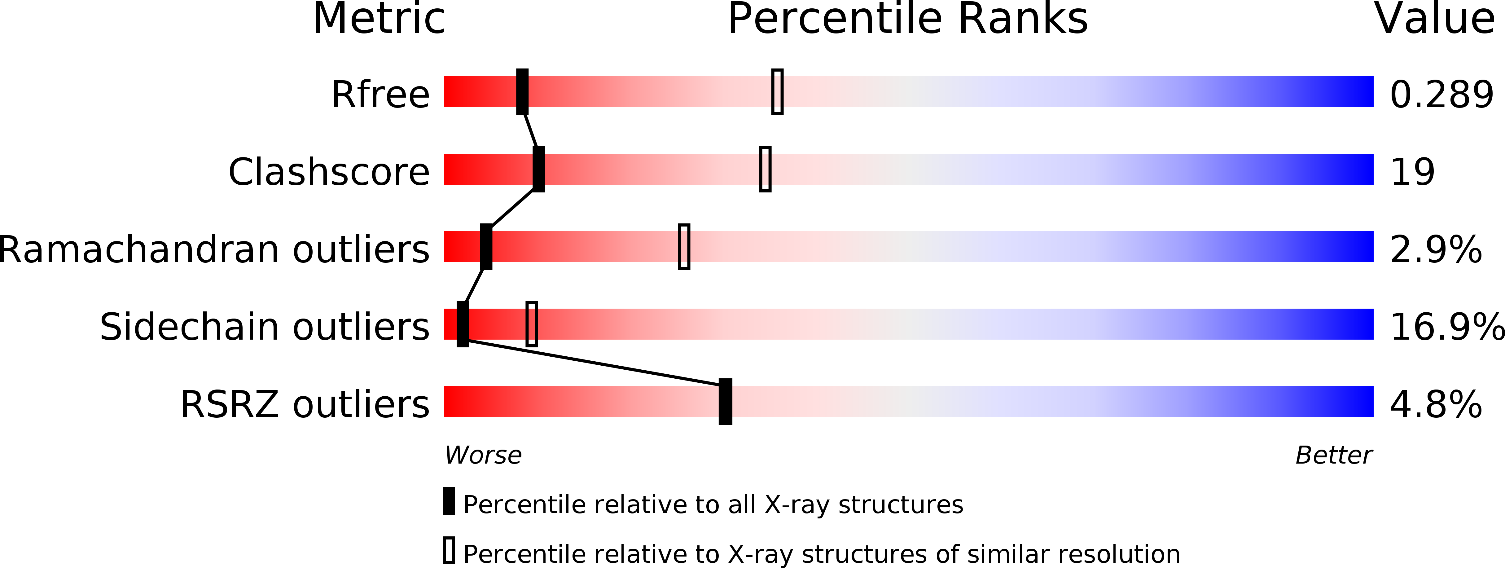
Resolution:
3.31 Å
R-Value Free:
0.29
R-Value Work:
0.26
R-Value Observed:
0.26
Space Group:
P 43