3d95 image
Deposition Date 2008-05-26
Release Date 2008-07-22
Last Version Date 2024-02-21
Entry Detail
PDB ID:
3D95
Title:
Crystal Structure of the R132K:Y134F:R111L:L121E:T54V Mutant of Apo-Cellular Retinoic Acid Binding Protein Type II at 1.20 Angstroms Resolution
Biological Source:
Source Organism:
Homo sapiens (Taxon ID: 9606)
Host Organism:
Method Details:
Experimental Method:
Resolution:
1.20 Å
R-Value Free:
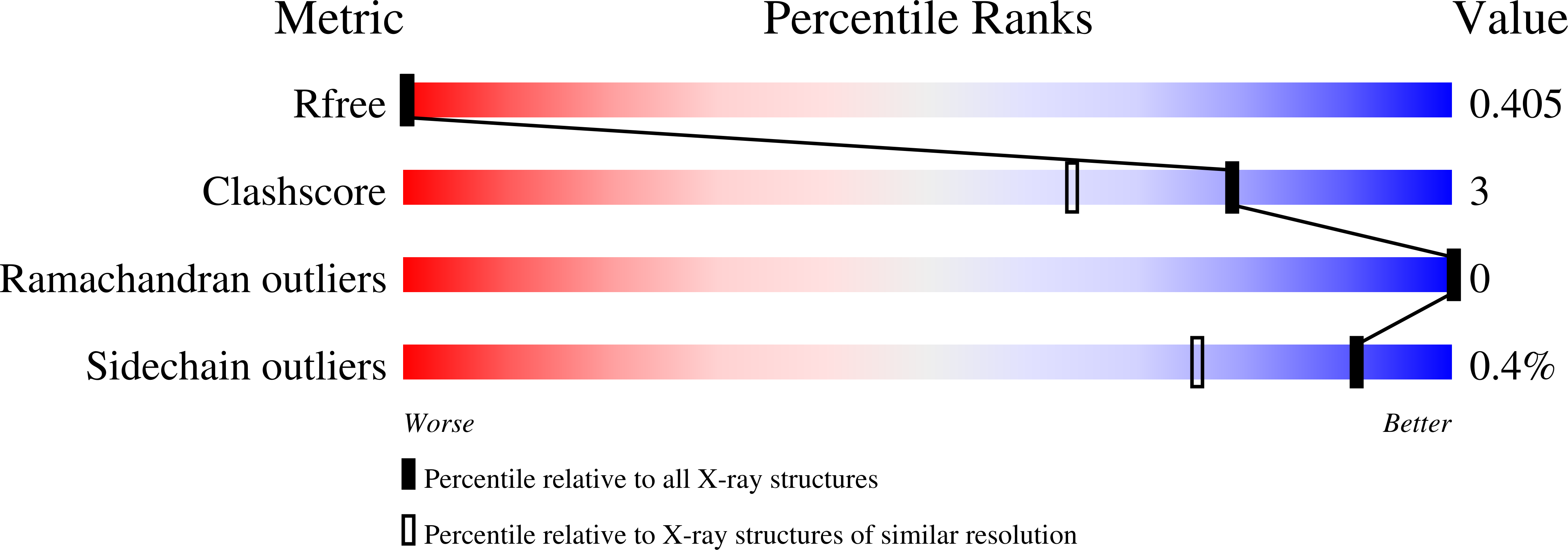
0.17
R-Value Work:
0.13
R-Value Observed:
0.14
Space Group:
P 1
Macromolecular Entities
Structures with similar UniProt ID
Protein Blast
Polymer Type:polypeptide(L)
Molecule:Cellular retinoic acid-binding protein 2
Gene (Uniprot):CRABP2
Mutagens:R132K, Y134F, R111L, L121E, T54V
Chain IDs:A, B
Chain Length:137
Number of Molecules:2
Biological Source:Homo sapiens
Primary Citation
Structural analysis of site-directed mutants of cellular retinoic acid-binding protein II addresses the relationship between structural integrity and ligand binding.
Acta Crystallogr.,Sect.D 64 1228 1239 (2008)
PMID: 19018099 DOI: 10.1107/S0907444908032216

Abstact

The structural integrity of cellular retinoic acid-binding protein II (CRABPII) has been investigated using the crystal structures of CRABPII mutants. The overall fold was well maintained by these CRABPII mutants, each of which carried multiple different mutations. A water-mediated network is found to be present across the large binding cavity, extending from Arg111 deep inside the cavity to the alpha2 helix at its entrance. This chain of interactions acts as a ;pillar' that maintains the integrity of the protein. The disruption of the water network upon loss of Arg111 leads to decreased structural integrity of the protein. A water-mediated network can be re-established by introducing the hydrophilic Glu121 inside the cavity, which results in a rigid protein with the alpha2 helix adopting an altered conformation compared with wild-type CRABPII.

Legend

Protein

Chemical

Disease

Primary Citation of related structures