Deposition Date
2008-01-25
Release Date
2008-09-23
Last Version Date
2024-10-30
Entry Detail
PDB ID:
3C2W
Keywords:
Title:
Crystal structure of the photosensory core domain of P. aeruginosa bacteriophytochrome PaBphP in the Pfr state
Biological Source:
Source Organism(s):
Pseudomonas aeruginosa (Taxon ID: 287)
Expression System(s):
Method Details:
Experimental Method:
Resolution:
2.90 Å
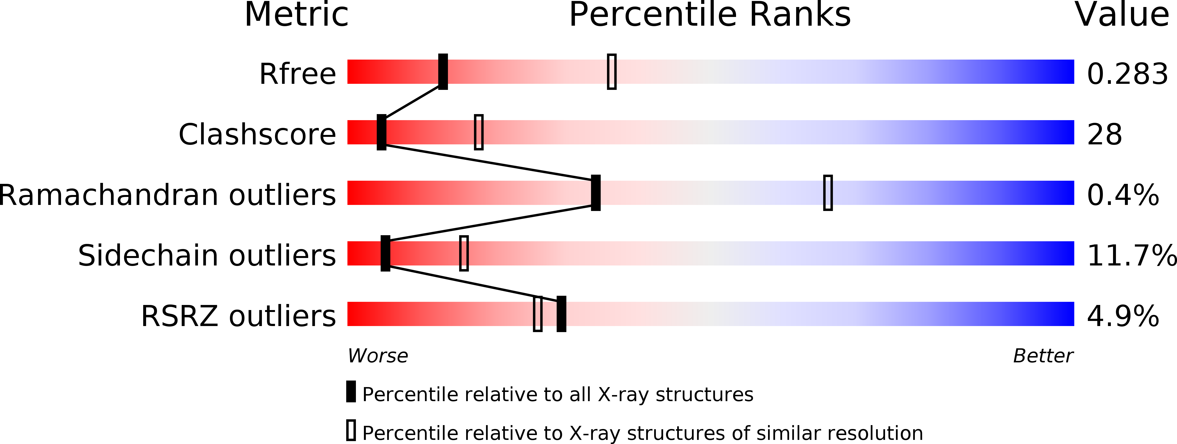
R-Value Free:
0.28
R-Value Work:
0.21
R-Value Observed:
0.22
Space Group:
C 2 2 21